We apologize for Proteopedia being slow to respond. For the past two years, a new implementation of Proteopedia has been being built. Soon, it will replace this 18-year old system. All existing content will be moved to the new system at a date that will be announced here.
Sandbox Reserved 1717
From Proteopedia
(Difference between revisions)
| Line 34: | Line 34: | ||
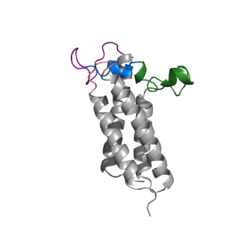
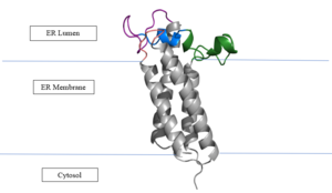
[[Image:VKORmembrane.png|300px|left|thumb|Figure 3. Orientation in Endoplasmic Reticulum: The cap region is partially oriented in the ER Lumen, however the active site remains within the ER membrane. The Beta Hairpin, Loop 3-4, Cap Loop are all in the ER Lumen. The Anchor is partially within the ER lumen, and partially embedded in the ER membrane. The anchor is what attaches the cap domain and stabilizes it, which allows the cap domain to cover the active site. ]] | [[Image:VKORmembrane.png|300px|left|thumb|Figure 3. Orientation in Endoplasmic Reticulum: The cap region is partially oriented in the ER Lumen, however the active site remains within the ER membrane. The Beta Hairpin, Loop 3-4, Cap Loop are all in the ER Lumen. The Anchor is partially within the ER lumen, and partially embedded in the ER membrane. The anchor is what attaches the cap domain and stabilizes it, which allows the cap domain to cover the active site. ]] | ||
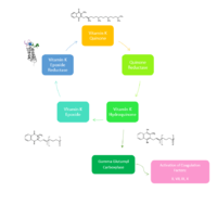
The reaction catalyzed by VKOR is a redox reaction. ''Vitamin K Epoxide => Vitamin K Quinone'' Vitamin K Epoxide is reduced by transferring two electrons through a disulfide bond. These disulfide bonds come from the conserved cysteines. This redox reaction that is catalyzed by VKOR produces Vitamin K Quinone. | The reaction catalyzed by VKOR is a redox reaction. ''Vitamin K Epoxide => Vitamin K Quinone'' Vitamin K Epoxide is reduced by transferring two electrons through a disulfide bond. These disulfide bonds come from the conserved cysteines. This redox reaction that is catalyzed by VKOR produces Vitamin K Quinone. | ||
| + | |||
| + | |||
| + | |||
| + | |||
| + | |||
| + | |||
| + | |||
| + | |||
| + | |||
| + | |||
| + | |||
| + | |||
| + | |||
| + | |||
| + | |||
| + | |||
| + | |||
| + | |||
| + | |||
| Line 49: | Line 68: | ||
== Vitamin K Epoxide == | == Vitamin K Epoxide == | ||
| - | [[Image:Vitamin K epoxide.jpg|500 px|right|thumb|Figure | + | [[Image:Vitamin K epoxide.jpg|500 px|right|thumb|Figure 4. Vitamin K Epoxide structure]] |
| - | As | + | As mentioned above, Vitamin K epoxide is a part of the Vitamin K cycle, necessary for blood coagulation. In the cycle, Vitamin K epoxide reductase (VKOR) reduces Vitamin K epoxide to quinone, or the active form of Vitamin K. What is occurring is VKOR donated electrons to Vitamin K epoxide, and those electrons come from the S-H of one of the cysteine pairs discussed above. The one cysteine pair has to be reduced for the transfer of electrons to the substrate can occur. |
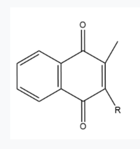
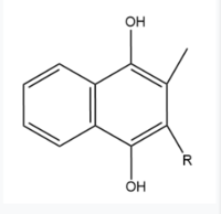
| + | Two other notable structures are Vitamin K Quinone (Fig. 5) and Vitamin K Hydroquinone (Fig. 6). Vitamin K Quinone is the product that is released after the reaction with Vitamin K Epoxide and VKOR. (Fig. 2) | ||
| + | [[Image:Vitaminkquinone.PNG|200 px|left|thumb|Figure 5. Vitamin K Quinone structure]] [[Image:Vitaminkhydroquinone.PNG|200 px|right|thumb|Figure 6. Vitamin K Hydroquinone structure]] | ||
=== Binding === | === Binding === | ||
| - | + | ||
| + | In its resting state, VKOR is in its open conformation. The Vitamin K epoxide enters through the isoprenyl-chain tunnel. The ketones on the VK epoxide bind to Asn80 and Tyr139 on VKOR. With Vitamin K epoxide bound, the cysteines of VKOR are partially oxidized, and concurrently reduce the substrate. A disulfide bond then forms between Cys51 and Cys132, resulting in the closed conformation. This leaves the sulfur on Cys43 and the sulfur on Cys135 protonated. The available hydrogens on these cysteines are utilized in reducing the epoxide. First, the free sulfur on Cys43 attacks Cys51 to form a new disulfide bond. With the loss of hydrogen from Cys43 in the formation of the new disulfide bond, an electron transfer is made to VKO. Next, the sulfur on Cys132 and the sulfur on Cys135 form a new disulfide bond. The hydrogen that was present on Cys135 is lost in the formation of the disulfide bond, allowing for an electron transfer to the oxygen of the epoxide. With these cysteine pairs formed, VKOR is left in an open conformation. The end products are Vitamin K/quinone and water. | ||
| Line 62: | Line 84: | ||
A set of four cysteines is consistently conserved in all VKOR homologs. In the human homolog (HsVKOR) these cysteines are Cys43, Cys51, Cys132, and Cys 135. <scene name='90/904321/Cysteines/6'>Significant Cysteines</scene> In the Pufferfish homolog (TrVKORL) these cysteines, due to Cryo-EM differences,are Cys52, Cys55, Cys141, and Cys144. These cysteines are the key factor that allow for Vitamin K Epoxide Reductase to perform its function, which is to open the epoxide ring on Vitamin K Epoxide in order to re-make Vitamin K Quinone. In the closed conformation, that is induced when Vitamin K binds in the hydrophobic pocket, Cys-132 binds to Cys-51 and Cys-135 will bind to the 3' hydroxyl group on Vitamin K Epoxide, which allows for the electron transfer to open up the epoxide ring. <scene name='90/904321/Cys52disulfidecys55/9'>Electron Transfer through Cysteine138 TrVKORL</scene> | A set of four cysteines is consistently conserved in all VKOR homologs. In the human homolog (HsVKOR) these cysteines are Cys43, Cys51, Cys132, and Cys 135. <scene name='90/904321/Cysteines/6'>Significant Cysteines</scene> In the Pufferfish homolog (TrVKORL) these cysteines, due to Cryo-EM differences,are Cys52, Cys55, Cys141, and Cys144. These cysteines are the key factor that allow for Vitamin K Epoxide Reductase to perform its function, which is to open the epoxide ring on Vitamin K Epoxide in order to re-make Vitamin K Quinone. In the closed conformation, that is induced when Vitamin K binds in the hydrophobic pocket, Cys-132 binds to Cys-51 and Cys-135 will bind to the 3' hydroxyl group on Vitamin K Epoxide, which allows for the electron transfer to open up the epoxide ring. <scene name='90/904321/Cys52disulfidecys55/9'>Electron Transfer through Cysteine138 TrVKORL</scene> | ||
| - | |||
| - | |||
== Warfarin == | == Warfarin == | ||
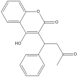
[https://en.wikipedia.org/wiki/Warfarin Warfarin] is the most common [https://en.wikipedia.org/wiki/Vitamin_K_antagonist Vitamin K antagonist (VKA)]. Warfarin is a competitive inhibitor, taking the place of Vitamin K Epoxide (VKO) in the active site of Vitamin K Epoxide Reductase (VKOR). When warfarin binds in the active site, it causes VKOR to go into the closed conformation. | [https://en.wikipedia.org/wiki/Warfarin Warfarin] is the most common [https://en.wikipedia.org/wiki/Vitamin_K_antagonist Vitamin K antagonist (VKA)]. Warfarin is a competitive inhibitor, taking the place of Vitamin K Epoxide (VKO) in the active site of Vitamin K Epoxide Reductase (VKOR). When warfarin binds in the active site, it causes VKOR to go into the closed conformation. | ||
| - | [[Image:warfarin.jpg|400 px|right|thumb|Figure | + | [[Image:warfarin.jpg|400 px|right|thumb|Figure 7. 2-Dimensional structure of Warfarin]] |
=== Binding === | === Binding === | ||
Revision as of 19:10, 5 April 2022
Vitamin K Epoxide Reductase
| |||||||||||
