This old version of Proteopedia is provided for student assignments while the new version is undergoing repairs. Content and edits done in this old version of Proteopedia after March 1, 2026 will eventually be lost when it is retired in about June of 2026.
Apply for new accounts at the new Proteopedia. Your logins will work in both the old and new versions.
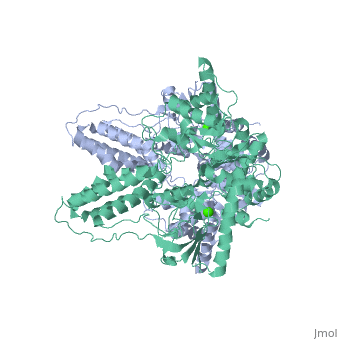
2wqd
From Proteopedia
(Difference between revisions)
| Line 3: | Line 3: | ||
<StructureSection load='2wqd' size='340' side='right'caption='[[2wqd]], [[Resolution|resolution]] 2.40Å' scene=''> | <StructureSection load='2wqd' size='340' side='right'caption='[[2wqd]], [[Resolution|resolution]] 2.40Å' scene=''> | ||
== Structural highlights == | == Structural highlights == | ||
| - | <table><tr><td colspan='2'>[[2wqd]] is a 1 chain structure with sequence from [https://en.wikipedia.org/wiki/ | + | <table><tr><td colspan='2'>[[2wqd]] is a 1 chain structure with sequence from [https://en.wikipedia.org/wiki/Staphylococcus_aureus Staphylococcus aureus]. Full crystallographic information is available from [http://oca.weizmann.ac.il/oca-bin/ocashort?id=2WQD OCA]. For a <b>guided tour on the structure components</b> use [https://proteopedia.org/fgij/fg.htm?mol=2WQD FirstGlance]. <br> |
| - | </td></tr><tr id='ligand'><td class="sblockLbl"><b>[[Ligand|Ligands:]]</b></td><td class="sblockDat" id="ligandDat"><scene name='pdbligand=CA:CALCIUM+ION'>CA</scene></td></tr> | + | </td></tr><tr id='method'><td class="sblockLbl"><b>[[Empirical_models|Method:]]</b></td><td class="sblockDat" id="methodDat">X-ray diffraction, [[Resolution|Resolution]] 2.4Å</td></tr> |
| + | <tr id='ligand'><td class="sblockLbl"><b>[[Ligand|Ligands:]]</b></td><td class="sblockDat" id="ligandDat"><scene name='pdbligand=CA:CALCIUM+ION'>CA</scene></td></tr> | ||
<tr id='resources'><td class="sblockLbl"><b>Resources:</b></td><td class="sblockDat"><span class='plainlinks'>[https://proteopedia.org/fgij/fg.htm?mol=2wqd FirstGlance], [http://oca.weizmann.ac.il/oca-bin/ocaids?id=2wqd OCA], [https://pdbe.org/2wqd PDBe], [https://www.rcsb.org/pdb/explore.do?structureId=2wqd RCSB], [https://www.ebi.ac.uk/pdbsum/2wqd PDBsum], [https://prosat.h-its.org/prosat/prosatexe?pdbcode=2wqd ProSAT]</span></td></tr> | <tr id='resources'><td class="sblockLbl"><b>Resources:</b></td><td class="sblockDat"><span class='plainlinks'>[https://proteopedia.org/fgij/fg.htm?mol=2wqd FirstGlance], [http://oca.weizmann.ac.il/oca-bin/ocaids?id=2wqd OCA], [https://pdbe.org/2wqd PDBe], [https://www.rcsb.org/pdb/explore.do?structureId=2wqd RCSB], [https://www.ebi.ac.uk/pdbsum/2wqd PDBsum], [https://prosat.h-its.org/prosat/prosatexe?pdbcode=2wqd ProSAT]</span></td></tr> | ||
</table> | </table> | ||
== Function == | == Function == | ||
| - | + | [https://www.uniprot.org/uniprot/PT1_STAAU PT1_STAAU] General (non sugar-specific) component of the phosphoenolpyruvate-dependent sugar phosphotransferase system (sugar PTS). This major carbohydrate active-transport system catalyzes the phosphorylation of incoming sugar substrates concomitantly with their translocation across the cell membrane. Enzyme I transfers the phosphoryl group from phosphoenolpyruvate (PEP) to the phosphoryl carrier protein (HPr). | |
== Evolutionary Conservation == | == Evolutionary Conservation == | ||
[[Image:Consurf_key_small.gif|200px|right]] | [[Image:Consurf_key_small.gif|200px|right]] | ||
| Line 36: | Line 37: | ||
</StructureSection> | </StructureSection> | ||
[[Category: Large Structures]] | [[Category: Large Structures]] | ||
| - | [[Category: Baumann | + | [[Category: Staphylococcus aureus]] |
| - | [[Category: Erni | + | [[Category: Baumann U]] |
| - | [[Category: Oberholzer | + | [[Category: Erni B]] |
| - | [[Category: Schneider | + | [[Category: Oberholzer AE]] |
| - | [[Category: Siebold | + | [[Category: Schneider P]] |
| - | + | [[Category: Siebold C]] | |
| - | + | ||
| - | + | ||
| - | + | ||
| - | + | ||
| - | + | ||
| - | + | ||
| - | + | ||
| - | + | ||
| - | + | ||
Current revision
Crystal structure of enzyme I of the phosphoenolpyruvate:sugar phosphotransferase system in the dephosphorylated state
| |||||||||||