We apologize for Proteopedia being slow to respond. For the past two years, a new implementation of Proteopedia has been being built. Soon, it will replace this 18-year old system. All existing content will be moved to the new system at a date that will be announced here.
Neurofibromin
From Proteopedia
(Difference between revisions)
| Line 3: | Line 3: | ||
Neurofibromin is a cytoplasmic protein located close to the cell membrane that is encoded by the ''NF1'' gene located on chromosome 17 <ref name= ''Bergoug''>PMID:33121128</ref>. It is a suppressor of the [http://https://www.cancer.gov/publications/dictionaries/cancer-terms/def/ras-gene-family Ras] oncogene through its effect on the rate of catalysis from <scene name='90/904325/Ras_full_structure/2'>Ras-GTP</scene> (active) to Ras-GDP (inactive)<ref name= ''Hall''>PMID:12213964</ref>. NF increasing the rate of catalysis of Ras means that Ras spends more time in its inactive state and cannot cause unnecessary cell proliferation linked to cancer<ref name= ''Cimino''>PMID:29478615</ref>. | Neurofibromin is a cytoplasmic protein located close to the cell membrane that is encoded by the ''NF1'' gene located on chromosome 17 <ref name= ''Bergoug''>PMID:33121128</ref>. It is a suppressor of the [http://https://www.cancer.gov/publications/dictionaries/cancer-terms/def/ras-gene-family Ras] oncogene through its effect on the rate of catalysis from <scene name='90/904325/Ras_full_structure/2'>Ras-GTP</scene> (active) to Ras-GDP (inactive)<ref name= ''Hall''>PMID:12213964</ref>. NF increasing the rate of catalysis of Ras means that Ras spends more time in its inactive state and cannot cause unnecessary cell proliferation linked to cancer<ref name= ''Cimino''>PMID:29478615</ref>. | ||
== Structure == | == Structure == | ||
| - | ===Important Structural Features=== | ||
| - | ====Active Site==== | ||
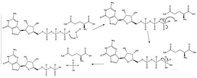
| - | The <scene name='90/904326/Active_site_with_residues/6'>active site</scene> for GTP hydrolysis of Ras is located in the Gap-related domain of neurofibromin. The catalytic residues include R68, Q61, and Y32, as well as magnesium and water molecules. Arginine is referred to as an [http://https://en.wikipedia.org/wiki/Arginine_finger “arginine finger”] because it points into the binding site of GTP to stabilize and orient the position of glutamine through a network of hydrogen bonds between water molecules. This arginine comes from the Gap-related domain of neurofibromin. When GDP is bound, glutamine is too far away to perform its catalytic action. Glutamine interacts with the gamma phosphate via a hydrogen bond created from an interaction between a water molecule and the gamma phosphate. When GTP is bound, tyrosine moves inward to face it. In the GDP bound form, tyrosine faces outward. | ||
====Conformations==== | ====Conformations==== | ||
| Line 17: | Line 14: | ||
==== CSRD and CTD ==== | ==== CSRD and CTD ==== | ||
The Cysteine-Serine-rich domain (CSRD) and C-terminal domain (CTD) contain phosphorylation sites. The CSRD is able to be phosphorylated by protein kinases A and C. Phosphorylation by protein kinase C is a positive regulator of neurofibromin activity. The CTD is phosphorylated primarily by protein kinase C. This domain is a negative regulator of neurofibromin activity if particular residues are phosphorylated. It also plays an important role in tubulin binding, as it helps in the transition from metaphase to anaphase. CTD contains a nuclear localization signal as well. | The Cysteine-Serine-rich domain (CSRD) and C-terminal domain (CTD) contain phosphorylation sites. The CSRD is able to be phosphorylated by protein kinases A and C. Phosphorylation by protein kinase C is a positive regulator of neurofibromin activity. The CTD is phosphorylated primarily by protein kinase C. This domain is a negative regulator of neurofibromin activity if particular residues are phosphorylated. It also plays an important role in tubulin binding, as it helps in the transition from metaphase to anaphase. CTD contains a nuclear localization signal as well. | ||
| + | ===Important Structural Features=== | ||
| + | ====Active Site==== | ||
| + | The <scene name='90/904326/Active_site_with_residues/6'>active site</scene> for GTP hydrolysis of Ras is located in the Gap-related domain of neurofibromin. The catalytic residues include R68, Q61, and Y32, as well as magnesium and water molecules. Arginine is referred to as an [http://https://en.wikipedia.org/wiki/Arginine_finger “arginine finger”] because it points into the binding site of GTP to stabilize and orient the position of glutamine through a network of hydrogen bonds between water molecules. This arginine comes from the Gap-related domain of neurofibromin. When GDP is bound, glutamine is too far away to perform its catalytic action. Glutamine interacts with the gamma phosphate via a hydrogen bond created from an interaction between a water molecule and the gamma phosphate. When GTP is bound, tyrosine moves inward to face it. In the GDP bound form, tyrosine faces outward. | ||
| + | |||
| + | new? | ||
| + | Ras and Neurofibromin associate through an arginine residue, 1276, that comes from neurofibromin. This arginine is referred to as the [http://https://en.wikipedia.org/wiki/Arginine_finger “arginine finger”] and assists in the hydrolysis of GTP by binding to a backbone carbon atom of tyrosine 32 of Ras when neurofibromin is in the open conformation. It points into the GTP binding site of Ras when neurofibromin is in the open conformation. R1276 also helps stabilize the position of Glutamine 61, a key catalytic residue, through hydrogen bonds. | ||
| + | |||
| + | Glutamine 61 of Ras is a residue that facilitates the conversion of GTP to GDP, turning Ras from its active state to inactive state. There is a catalytic water molecule that glutamine interacts with to position the molecule for a nucleophilic attack on the gamma phosphate of GTP. Mutations of this residue have been related to lower rates of hydrolysis. <ref name= ''Frech''>PMID:8136358</ref>. Tyrosine 32 makes water-mediated hydrogen bonds with the gamma phosphate of GTP. This position is also where Ras is phosphorylation to promote the activity of GTPase-activating proteins and GTP hydrolysis. <ref name= ''Bunda''>DOI:10.1038/ncomms9859</ref> | ||
==RAS Complex== | ==RAS Complex== | ||
===Mechanism of Ras Coupled with Neurofibromin=== | ===Mechanism of Ras Coupled with Neurofibromin=== | ||
Revision as of 18:05, 7 April 2022
| |||||||||||
References
- ↑ Bergoug M, Doudeau M, Godin F, Mosrin C, Vallee B, Benedetti H. Neurofibromin Structure, Functions and Regulation. Cells. 2020 Oct 27;9(11). pii: cells9112365. doi: 10.3390/cells9112365. PMID:33121128 doi:http://dx.doi.org/10.3390/cells9112365
- ↑ Hall BE, Bar-Sagi D, Nassar N. The structural basis for the transition from Ras-GTP to Ras-GDP. Proc Natl Acad Sci U S A. 2002 Sep 17;99(19):12138-42. Epub 2002 Sep 4. PMID:12213964 doi:http://dx.doi.org/10.1073/pnas.192453199
- ↑ Cimino PJ, Gutmann DH. Neurofibromatosis type 1. Handb Clin Neurol. 2018;148:799-811. doi: 10.1016/B978-0-444-64076-5.00051-X. PMID:29478615 doi:http://dx.doi.org/10.1016/B978-0-444-64076-5.00051-X
- ↑ Frech M, Darden TA, Pedersen LG, Foley CK, Charifson PS, Anderson MW, Wittinghofer A. Role of glutamine-61 in the hydrolysis of GTP by p21H-ras: an experimental and theoretical study. Biochemistry. 1994 Mar 22;33(11):3237-44. doi: 10.1021/bi00177a014. PMID:8136358 doi:http://dx.doi.org/10.1021/bi00177a014
- ↑ Bunda S, Burrell K, Heir P, Zeng L, Alamsahebpour A, Kano Y, Raught B, Zhang ZY, Zadeh G, Ohh M. Inhibition of SHP2-mediated dephosphorylation of Ras suppresses oncogenesis. Nat Commun. 2015 Nov 30;6:8859. doi: 10.1038/ncomms9859. PMID:26617336 doi:http://dx.doi.org/10.1038/ncomms9859
- ↑ Scheffzek K, Shivalingaiah G. Ras-Specific GTPase-Activating Proteins-Structures, Mechanisms, and Interactions. Cold Spring Harb Perspect Med. 2019 Mar 1;9(3). pii: cshperspect.a031500. doi:, 10.1101/cshperspect.a031500. PMID:30104198 doi:http://dx.doi.org/10.1101/cshperspect.a031500
- ↑ Prive GG, Milburn MV, Tong L, de Vos AM, Yamaizumi Z, Nishimura S, Kim SH. X-ray crystal structures of transforming p21 ras mutants suggest a transition-state stabilization mechanism for GTP hydrolysis. Proc Natl Acad Sci U S A. 1992 Apr 15;89(8):3649-53. doi: 10.1073/pnas.89.8.3649. PMID:1565661 doi:http://dx.doi.org/10.1073/pnas.89.8.3649
- ↑ Lupton CJ, Bayly-Jones C, D'Andrea L, Huang C, Schittenhelm RB, Venugopal H, Whisstock JC, Halls ML, Ellisdon AM. The cryo-EM structure of the human neurofibromin dimer reveals the molecular basis for neurofibromatosis type 1. Nat Struct Mol Biol. 2021 Dec;28(12):982-988. doi: 10.1038/s41594-021-00687-2., Epub 2021 Dec 9. PMID:34887559 doi:http://dx.doi.org/10.1038/s41594-021-00687-2
- ↑ Cimino PJ, Gutmann DH. Neurofibromatosis type 1. Handb Clin Neurol. 2018;148:799-811. doi: 10.1016/B978-0-444-64076-5.00051-X. PMID:29478615 doi:http://dx.doi.org/10.1016/B978-0-444-64076-5.00051-X
- ↑ Ly KI, Blakeley JO. The Diagnosis and Management of Neurofibromatosis Type 1. Med Clin North Am. 2019 Nov;103(6):1035-1054. doi: 10.1016/j.mcna.2019.07.004. PMID:31582003 doi:http://dx.doi.org/10.1016/j.mcna.2019.07.004
Proteopedia Page Contributors and Editors (what is this?)
Jordyn K. Lenard, Ryan D. Adkins, Michal Harel, OCA, Jaime Prilusky


