We apologize for Proteopedia being slow to respond. For the past two years, a new implementation of Proteopedia has been being built. Soon, it will replace this 18-year old system. All existing content will be moved to the new system at a date that will be announced here.
Sandbox Reserved 1716
From Proteopedia
(Difference between revisions)
| Line 32: | Line 32: | ||
== Vitamin K Epoxide == | == Vitamin K Epoxide == | ||
| - | [[Image:Vitamin K epoxide.jpg|500 px|right|thumb|Figure | + | [[Image:Vitamin K epoxide.jpg|500 px|right|thumb|Figure 4. Vitamin K Epoxide structure]] |
As mentioned above, Vitamin K epoxide is a component of the Vitamin K cycle and required for blood coagulation. In the cycle, VKOR reduces Vitamin K epoxide to Vitamin K Quinone, or the active form of Vitamin K. In this conversion, VKOR donates electrons to Vitamin K epoxide from the S-H of the active pair of cysteines, C132-C135. The mediated cysteine pair, C43-C51, has to be reduced for the transfer of electrons to the substrate to occur. | As mentioned above, Vitamin K epoxide is a component of the Vitamin K cycle and required for blood coagulation. In the cycle, VKOR reduces Vitamin K epoxide to Vitamin K Quinone, or the active form of Vitamin K. In this conversion, VKOR donates electrons to Vitamin K epoxide from the S-H of the active pair of cysteines, C132-C135. The mediated cysteine pair, C43-C51, has to be reduced for the transfer of electrons to the substrate to occur. | ||
| Line 38: | Line 38: | ||
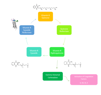
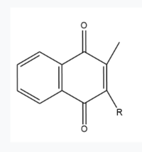
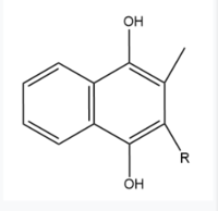
Two other notable structures from the Vitamin K cycle are Vitamin K Quinone (Fig. 5) and Vitamin K Hydroquinone (Fig. 6). Vitamin K Quinone is the product that is released after the reaction with Vitamin K Epoxide and VKOR. (Fig. 2) | Two other notable structures from the Vitamin K cycle are Vitamin K Quinone (Fig. 5) and Vitamin K Hydroquinone (Fig. 6). Vitamin K Quinone is the product that is released after the reaction with Vitamin K Epoxide and VKOR. (Fig. 2) | ||
| - | [[Image:Vitaminkquinone.PNG|200 px|left|thumb|Figure 3. Vitamin K Quinone structure]] [[Image:Vitaminkhydroquinone.PNG|200 px|right|thumb|Figure 4. Vitamin K Hydroquinone structure]] | ||
| - | === Binding === | ||
| + | [[Image:Vitaminkquinone.PNG|200 px|left|thumb|Figure 2. Vitamin K Quinone structure]] [[Image:Vitaminkhydroquinone.PNG|200 px|right|thumb|Figure 3. Vitamin K Hydroquinone structure]] | ||
| - | In its resting state, VKOR is in its <scene name='90/904322/Open_conformation/1'>open conformation</scene>. The Vitamin K epoxide enters through the isoprenyl-chain tunnel. The ketones on the VK epoxide bind to <scene name='90/904322/Vko_binding/1'>Asn80 and Tyr139</scene> on VKOR. With Vitamin K epoxide bound, the cysteines of VKOR are partially oxidized, and concurrently reduce the substrate. A disulfide bond then forms between Cys51 and Cys132, resulting in the closed conformation. This leaves the sulfur on Cys43 and the sulfur on Cys135 protonated. The available hydrogens on these cysteines are utilized in reducing the epoxide. First, the free sulfur on Cys43 attacks Cys51 to form a new disulfide bond. With the loss of hydrogen from Cys43 in the formation of the new disulfide bond, an electron transfer is made to VKO. Next, the sulfur on Cys132 and the sulfur on Cys135 form a new disulfide bond. The hydrogen that was present on Cys135 is lost in the formation of the disulfide bond, allowing for an electron transfer to the oxygen of the epoxide. With these cysteine pairs formed, VKOR is left in an open conformation. The end products are Vitamin K | + | |
| + | === Binding === | ||
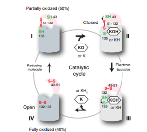
| + | In its resting state, VKOR is in its <scene name='90/904322/Open_conformation/1'>open conformation</scene>. The Vitamin K epoxide enters through the <scene name='90/904322/Tunnel/6'>isoprenyl-chain tunnel</scene>. The tunnel is located between TM2 (dark) and TM3 (light). The ketones on the VK epoxide bind to <scene name='90/904322/Vko_binding/1'>Asn80 and Tyr139</scene> on VKOR. With Vitamin K epoxide bound, the cysteines of VKOR are partially oxidized, and concurrently reduce the substrate. A disulfide bond then forms between Cys51 and Cys132, resulting in the closed conformation. This leaves the sulfur on Cys43 and the sulfur on Cys135 protonated. The available hydrogens on these cysteines are utilized in reducing the epoxide. First, the free sulfur on Cys43 attacks Cys51 to form a new disulfide bond. With the loss of hydrogen from Cys43 in the formation of the new disulfide bond, an electron transfer is made to VKO. Next, the sulfur on Cys132 and the sulfur on Cys135 form a new disulfide bond. The hydrogen that was present on Cys135 is lost in the formation of the disulfide bond, allowing for an electron transfer to the oxygen of the epoxide. With these cysteine pairs formed, VKOR is left in an open conformation. The end products are Vitamin K Quinone and water. | ||
==Catalytic Cycle== | ==Catalytic Cycle== | ||
| - | [[Image:VKORcycle.PNG|300px|right|thumb|'''Figure | + | [[Image:VKORcycle.PNG|300px|right|thumb|'''Figure 4: The catalytic cycle of Vitamin K Epoxide Reductase''' <ref name=”Shixuan”>PMID:33154105</ref> ]] |
| Line 68: | Line 69: | ||
| - | == | + | == Warfarin == |
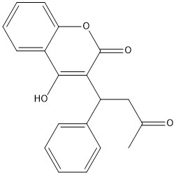
[https://en.wikipedia.org/wiki/Warfarin Warfarin] is the most common [https://en.wikipedia.org/wiki/Vitamin_K_antagonist Vitamin K antagonist (VKA)]. Warfarin is a competitive inhibitor, taking the place of Vitamin K Epoxide (VKO) in the active site of Vitamin K Epoxide Reductase (VKOR). When warfarin binds in the active site, it causes VKOR to go into the closed conformation. | [https://en.wikipedia.org/wiki/Warfarin Warfarin] is the most common [https://en.wikipedia.org/wiki/Vitamin_K_antagonist Vitamin K antagonist (VKA)]. Warfarin is a competitive inhibitor, taking the place of Vitamin K Epoxide (VKO) in the active site of Vitamin K Epoxide Reductase (VKOR). When warfarin binds in the active site, it causes VKOR to go into the closed conformation. | ||
| - | [[Image:warfarin.jpg|400 px|right|thumb|Figure | + | [[Image:warfarin.jpg|400 px|right|thumb|Figure 7. 2-Dimensional structure of Warfarin]] |
=== Binding === | === Binding === | ||
| - | |||
Warfarin also forms hydrogen bonds with <scene name='90/904322/Tyr_asn_binding_warfarin/2'>Asn80 and Tyr139</scene>. The specific bonds are between Asn80 and the 2-ketone group of warfarin and Tyr139 with the 4-hydroxyl group of warfarin. The rest of the warfarin interaction is determined by hydrophobic interactions. These hydrogen bonds provide the necessary recognition for the ligand to bind to the binding site to VKOR. | Warfarin also forms hydrogen bonds with <scene name='90/904322/Tyr_asn_binding_warfarin/2'>Asn80 and Tyr139</scene>. The specific bonds are between Asn80 and the 2-ketone group of warfarin and Tyr139 with the 4-hydroxyl group of warfarin. The rest of the warfarin interaction is determined by hydrophobic interactions. These hydrogen bonds provide the necessary recognition for the ligand to bind to the binding site to VKOR. | ||
| - | Warfarin binds with a slight difference compared to VKO. Warfarin binds at a slightly different angle. This creates a difference in how the cap loop and anchor domain interact and that affects the placement of <scene name='90/904322/Arg58/ | + | Warfarin binds with a slight difference compared to VKO. Warfarin binds at a slightly different angle. This creates a difference in how the cap loop and anchor domain interact and that affects the placement of <scene name='90/904322/Arg58/4'>Arg58</scene>. With VKO, Arg58 from the cap loop directly interacts with <scene name='90/904322/Arg58_vko/5'>Glu67</scene>. However, when warfarin binds, Arg58 is inserted between <scene name='90/904322/Arg58_warfarin/2'>Glu67 and His68</scene> of the anchor domain. |
| + | |||
| + | |||
=== Disease === | === Disease === | ||
| - | Vitamin K | + | [https://en.wikipedia.org/wiki/Vitamin_K_antagonist#:~:text=Vitamin%20K%20antagonists%20(VKA)%20are,the%20recycling%20of%20vitamin%20K. Vitamin K Antagonists] are first line therapeutics in the treatment of thromboembolic diseases, like a stroke or heart attack. <ref name=”Goy”>PMID:23034830<ref/>[https://en.wikipedia.org/wiki/Warfarin Warfarin] is the most common medication for this treatment, acting as a blood thinner. Warfarin binding to VKOR prevents the triggering of coagulation factors that form blood clots. |
== References == | == References == | ||
| + | <references/> | ||
| + | |||
| + | |||
| + | |||
| + | |||
| + | |||
| + | (DON’T INCLUDE) | ||
<ref name="Goodstadt">PMID:15276181</ref> Goodstadt, L., & Ponting, C. P. (2004). Vitamin K epoxide reductase: homology, active site and catalytic mechanism. ''Trends in biochemical sciences, 29''(6), 289–292. https://doi.org/10.1016/j.tibs.2004.04.004 | <ref name="Goodstadt">PMID:15276181</ref> Goodstadt, L., & Ponting, C. P. (2004). Vitamin K epoxide reductase: homology, active site and catalytic mechanism. ''Trends in biochemical sciences, 29''(6), 289–292. https://doi.org/10.1016/j.tibs.2004.04.004 | ||
Revision as of 16:20, 15 April 2022
Vitamin K Epoxide Reductase
| |||||||||||
