Sandbox Reserved 1709
From Proteopedia
| Line 17: | Line 17: | ||
=== Cap Domain === | === Cap Domain === | ||
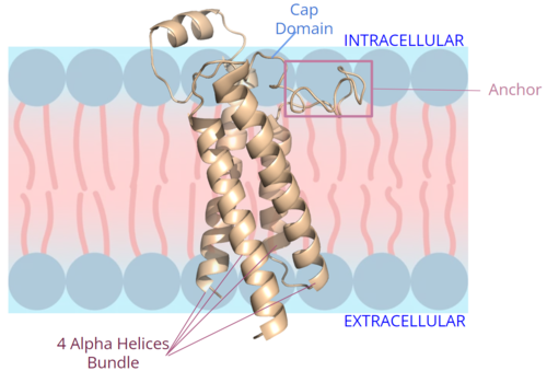
[[Image:New membrane pic bundle.png|500 px|right|thumb|Figure 2. Orientation and interactions of the VKOR components cap domain, anchor domain, and helical tunnel within the cell membrane.]] | [[Image:New membrane pic bundle.png|500 px|right|thumb|Figure 2. Orientation and interactions of the VKOR components cap domain, anchor domain, and helical tunnel within the cell membrane.]] | ||
| - | A key part of VKOR is the function of the <scene name='90/904314/Cap_domain/ | + | A key part of VKOR is the function of the <scene name='90/904314/Cap_domain/8'>cap domain</scene>, which is located right above the helices of VKOR towards the intracellular part of the membrane. The cap has a helical shape and is located in close proximity to two other domains: the Anchor domain and beta hairpin. This combination of domains help to maintain the proper orientation in the membrane. The cap domain assists with activating Vitamin K as it induces the structural change of VKOR from the open conformation to the closed conformation upon substrate binding. Cap rearrangement and transition to the closed conformation initiates a domino effect through the [https://reader.elsevier.com/reader/sd/pii/S0021925820001386?token=9F8E1964241D20488CA55E035D35D9A5D650A7B3FDAD9A5579598A8DC00127539BE71CF1785B117102144AC1F41ABB6C&originRegion=us-east-1&originCreation=20220329001707/ catalytic mechanism]. The cap domain has critical interactions that stabilize the closed conformation including a <scene name='90/904314/Disulfide_bridge_stabilization/8'>disulfide bridge</scene> between C43 and C51, and polar interactions from D44. These interactions are broken up by reactive cysteines to induce different conformations and help facilitate this transition from the open conformation to the closed conformation during the activation of Vitamin K. |
=== Anchor === | === Anchor === | ||
| - | The <scene name='90/904314/Anchor_domain/ | + | The <scene name='90/904314/Anchor_domain/12'>anchor domain</scene> is a key part of the VKOR structure and function that protrudes from the side of VKOR with the primary role of stabilizing the enzyme within the membrane. It sits on top of the membrane surface, as shown in figure 2, such that anchor residues can interact with the cell membrane to maintain proper proximity for VKOR activity. To accomplish this, hydrophilic residues are positioned to interact with the outer hydrophilic leaflet of the bilipid membrane, while the hydrophobic residues on the anchor have strong interactions with the inner hydrophobic leaflet of the bilipid membrane. These <scene name='90/904314/Anchor_domain/13'>polar and nonpolar interactions</scene> allow for VKOR to remain in the proper membrane arrangement and proximity for Vitamin K to bind and be activated via the cap domain and active site. The anchor also serves a role in connecting the cap domain to the rest of the membrane so that it stabilizes its covering of the central binding pocket to keep the substrate within the active site during its catalytic activation. These membrane interactions allow for VKOR to stabilize in the membrane for proper activation of Vitamin K and catalytic function of the enzyme. |
==Catalytic Mechanism of VKOR== | ==Catalytic Mechanism of VKOR== | ||
| Line 30: | Line 30: | ||
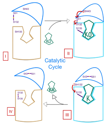
[[Image:Ss_of_catalytic_mech.png|350 px| right| thumb | Figure 3. Mechanism of VKOR.]] | [[Image:Ss_of_catalytic_mech.png|350 px| right| thumb | Figure 3. Mechanism of VKOR.]] | ||
| - | The catalytic mechanism of VKOR is highly regulated and use <scene name='90/904314/Stage_4_catalytic_cycle/ | + | The catalytic mechanism of VKOR is highly regulated and use <scene name='90/904314/Stage_4_catalytic_cycle/17'>four catalytic cysteine residues</scene> to activate Vitamin K necessary for blood coagulation. Figure 3 highlights these reactions that allow the substrate to be catalyzed to its active form through a series of 4 stages. The enzyme begins in <scene name='90/904314/Vkor_with_ko/1'>stage I</scene> in the open conformation with the cap domain open to allow substrate binding. Once a substrate binds, the cap domain transitions to the closed conformation when the C51-C132 disulfide bridge is attacked by reactive C43 located within the cap domain. This reaction forms a new disulfide bridge between C43 and C51 that pulls the cap domain over the binding pocket with the substrate bound to stabilize the closed conformation of VKOR. VKOR is now in <scene name='90/904314/Stage_2_catalytic_cycle/2'>stage II</scene>. Free cysteines are now available that provide strong stabilization of the closed conformation through interactions with the cap domain and the bound substrate. This puts the enzyme in <scene name='90/904314/Stage_3_catalytic_cycle/8'>stage III</scene>, where a free C135 is purposed to interact with the substrate within the binding pocket to stabilize it during activation. The catalytic free C132 located between the cap domain and helical tunnel is very reactive and will attack this C135 to break that interaction with the substrate and release the activated Vitamin K product into the blood stream to promote coagulation. Two very stable disulfide bridges between C43-C41 and C132-C135 are now present and VKOR is unbound, so the enzyme is in its final, unreactive <scene name='90/904314/Stage_4_catalytic_cycle/15'>stage IV</scene>. VKOR must undergo conformational changes to return to Stage 1 and reactivate its catalytic cysteines so that another molecule of Vitamin K can bind and be activated. |
== Disease and Treatment == | == Disease and Treatment == | ||
Revision as of 17:27, 18 April 2022
Vitamin K Epoxide Reductase
| |||||||||||
References
1. DJin, Da-Yun, Tie, Jian-Ke, and Stafford, Darrel W. "The Conversion of Vitamin K Epoxide to Vitamin K Quinone and Vitamin K Quinone to Vitamin K Hydroquinone Uses the Same Active Site Cysteines." Biochemistry 2007 46 (24), 7279-7283 [1].
2. Elshaikh, A. O., Shah, L., Joy Mathew, C., Lee, R., Jose, M. T., & Cancarevic, I. "Influence of Vitamin K on Bone Mineral Density and Osteoporosis" (2020) Cureus, 12(10), e10816. [2]
3. Guomin Shen, Weidong Cui, Qing Cao, Meng Gao, Hongli Liu, Gaigai Su, Michael L. Gross, Weikai Li. The catalytic mechanism of vitamin K epoxide reduction in a cellular environment. (2021) Journal of Biological Chemistry, Volume 296,100145. https://doi.org/10.1074/jbc.RA120.015401.
4. Li, Weikai et al. “Structure of a bacterial homologue of vitamin K epoxide reductase.” Nature vol. 463,7280 (2010): 507-12. doi:10.1038/nature08720.
5. Liu S, Li S, Shen G, Sukumar N, Krezel AM, Li W. Structural basis of antagonizing the vitamin K catalytic cycle for anticoagulation. Science. 2021 Jan 1;371(6524):eabc5667. doi: 10.1126/science.abc5667. Epub 2020 Nov 5. PMID: 33154105; PMCID: PMC7946407.
6. Patel S, Singh R, Preuss CV, Patel N. Warfarin. 2022 Jan 19. In: StatPearls [Internet]. Treasure Island (FL): StatPearls Publishing; 2022 Jan–. PMID: 29261922.
7. Yang W., et. al. “VKORC1 Haplotypes Are Associated With Arterial Vascular Diseases (Stroke, Coronary Heart Disease, and Aortic Dissection)” (2006) Circulation. ;113:1615–1621 [3]


