|
This page describes the use of FirstGlance in Jmol version 4.0. Its release is expected soon, but it is not yet publicly available. You can test this near-final version which is also used in the FirstGlance links below, but if you upload a file it will revert to the older version of FirstGlance.
|
Virus capsids and similarly large protein assemblies can be conveniently visualized and analyzed with FirstGlance in Jmol. Below some examples are explained and illustrated, but here is a quick start: polio virus capsid in FirstGlance (1pov).
FirstGlance in Jmol automatically constructs biological unit 1, thought to be the major functional quaternary assembly. Biological unit 1 is shown initially by default, and you can start separate sessions to show the asymmetric unit or other biological units when more than one are specified. When the resulting assembly is too large to work smoothly and efficiently in FirstGlance in Jmol (all Javascript), FirstGlance will automatically simplify the model to alpha carbons, or when necessary, to a subset of alpha carbons.
Polio Virus
Polio virus is small (among viruses pathogenic for humans) with an RNA genome inside a single-shell protein capsid. 1pov
made up from 3 proteins, 60 copies of each of VP0, VP1 and VP3 (from proteolysis of polyprotein UniProt P03300). 1pov provides the structure of the 3 chains in the asymmetric unit, along with instructions for constructing the capsid (Biological Unit 1).
The polio capsid has an icosahedral construction with 12 vertices defining 20 triangular faces. Notice that the larger blue mesas protruding on the surface have 5-fold symmetry. These are pentagonal capsomeres at the 12 vertices. The capsid is composed of 180 protein chains totaling about 377K non-H atoms.
Quick Start: polio virus capsid in FirstGlance
Measuring Capsid Diameter
FirstGlance/How To Measure A Virus Capsid provides step by step instructions on how to view a slab of the capsid and measure distances.
Color Schemes
(see caption). In addition to coloring by distance from the center, or assigning a distinct color to each group of sequence-identical chains, FirstGlance offers coloring by amino to carboxy rainbow, hydrophobic vs. polar, charge, and as illustrated below, evolutionary conservation.
Hide and Isolate
Hide and Isolate tools are provided in the Focus Box of FirstGlance. Clicking on a chain offers to hide or isolate all sequence-identical chains. Here,
Similarly one can easily isolate
or
Eastern Equine Encephalitis Virus
The (EEEV) consists of 240 copies of each of three distinct protein sequences, totaling 720 chains. It has nearly 2 million non-hydrogen atoms (so about 3.8 million atoms including hydrogen). The structure 6mx4 determined by electron microscopy (4.4 Å resolution) has 12 chains, with instructions for constructing the entire capsid using 60 copies of the asymmetric unit[3].
Analyze the EEEV capsid 6mx4 in FirstGlance
Double-Shelled Capsid
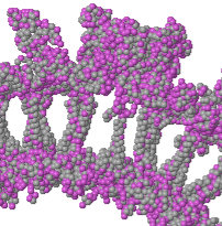
Immediately you can see that this is a double-layered capsid, with outer and inner shells. Between the shells are transmembrane alpha helical "spikes" or "posts" embedded in a lipid bilayer (not represented by any atoms here).
FirstGlance/How To Measure A Virus Capsid provides step by step instructions on how to view a slab of the capsid and measure distances.
The inside diameter is about 315 Å. The length of the transmembrane spikes/posts is about 35 Å, consistent with the thickness of a lipid bilayer.
(total 720 protein chains).
Transmembrane Posts
When all ~242K alpha carbons (rather than every 10th) are displayed in FirstGlance, performance is sluggish,
but more color schemes can be meaningfully applied.
Coloring alpha carbons in a slab by Hydrophobic, Polar reveals that the transmembrane posts are largely hydrophobic.
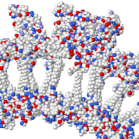
So, as expected, coloring the slab alpha carbons by charge
Anionic (-) / Cationic (+)
shows the posts to be nearly devoid of charges.
Clathrin Coat
This (3iyv), determined by electron microscopy at 7.9 Å resolution[1], represents the clathrin lattice structure enveloping coated pits and coated vesicles. The deposited model contains only alpha carbons. It is about 725 by 665 Å. There are 184K alpha carbons, so it would have about three million atoms including hydrogens, with a molecular mass of about 21 MDa. 96% of the model is composed of 108 clathrin heavy chains, each of which models 1,630 of the 1,675 residues in UniProt P49951. , each modeled as 70 of 243 residues of UniProt P04973.
Amino and Carboxy Termini
with its amino terminus blue and carboxy terminus red (with a spectal rainbow color sequence in between). When , it becomes evident that the carboxy termini are on the outside surface, while the amino termini form the inner surface.
Secondary Structure
Alpha Helices, Beta Strands , Loops
The amino terminal domain is nearly all all beta sheets and loops, arranged as seven 4-beta-strand sheets in a donut, forming a compact domain of 330 amino acids about 45 Å in diameter. The remaining 1,300 amino acids form a "snake" about 460 Å long and roughly 20 Å thick. It is nearly all antiparallel alpha helices orthogonal to the long dimension. When the entire clathrin coat model is colored by secondary structure, the outside surface is made of the alpha-helical snakes, and the inner surface is lined with the amino-terminal beta-sheet donuts.
Vault Nanocompartment
occurs in eukaryotes.
It is about 650 Å long and 380 Å wide.
Its function is unknown[2]. The vault is composed of 78 copies of a single protein sequence.
This model has nearly one million atoms (including hydrogens), and 61K alpha carbons.
Analyze the Simplified Vault in FirstGlance
Evolutionary Conservation

Vault with Conservation in FirstGlance (all 61K alpha carbons)
Conservation analysis was performed by ConSurf. In order for the pattern to be meaningful, all 61K alpha carbons were loaded into FirstGlance, which then applied the conservation colors.
Secondary Structure
amino-terminal portion consists of
a series of small antiparallel beta sheet domains. A long alpha helix forms the carboxy-terminal portion.
Alpha Helices, Beta Strands , Loops
Take a closer look at the or the .
Here is the
Bacterial Gas Vesicle
(In preparation.)


