This old version of Proteopedia is provided for student assignments while the new version is undergoing repairs. Content and edits done in this old version of Proteopedia after March 1, 2026 will eventually be lost when it is retired in about June of 2026.
Apply for new accounts at the new Proteopedia. Your logins will work in both the old and new versions.
1j3h
From Proteopedia
| Line 1: | Line 1: | ||
| - | [[Image:1j3h. | + | {{Seed}} |
| + | [[Image:1j3h.png|left|200px]] | ||
<!-- | <!-- | ||
| Line 9: | Line 10: | ||
{{STRUCTURE_1j3h| PDB=1j3h | SCENE= }} | {{STRUCTURE_1j3h| PDB=1j3h | SCENE= }} | ||
| - | + | ===Crystal structure of apoenzyme cAMP-dependent protein kinase catalytic subunit=== | |
| - | + | <!-- | |
| - | + | The line below this paragraph, {{ABSTRACT_PUBMED_12614615}}, adds the Publication Abstract to the page | |
| + | (as it appears on PubMed at http://www.pubmed.gov), where 12614615 is the PubMed ID number. | ||
| + | --> | ||
| + | {{ABSTRACT_PUBMED_12614615}} | ||
==About this Structure== | ==About this Structure== | ||
| Line 35: | Line 39: | ||
[[Category: Open conformation]] | [[Category: Open conformation]] | ||
[[Category: Preformed active site]] | [[Category: Preformed active site]] | ||
| - | ''Page seeded by [http://oca.weizmann.ac.il/oca OCA ] on | + | |
| + | ''Page seeded by [http://oca.weizmann.ac.il/oca OCA ] on Tue Jul 1 14:26:45 2008'' | ||
Revision as of 11:26, 1 July 2008
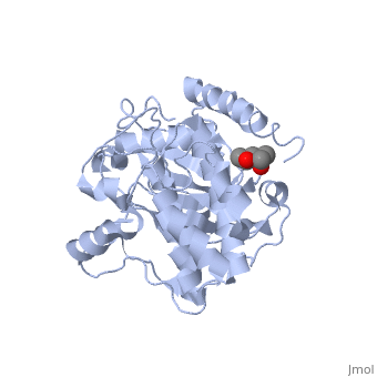

Crystal structure of apoenzyme cAMP-dependent protein kinase catalytic subunit
Template:ABSTRACT PUBMED 12614615
About this Structure
1J3H is a Single protein structure of sequence from Mus musculus. Full crystallographic information is available from OCA.
Reference
Dynamic features of cAMP-dependent protein kinase revealed by apoenzyme crystal structure., Akamine P, Madhusudan, Wu J, Xuong NH, Ten Eyck LF, Taylor SS, J Mol Biol. 2003 Mar 14;327(1):159-71. PMID:12614615
Page seeded by OCA on Tue Jul 1 14:26:45 2008
