We apologize for Proteopedia being slow to respond. For the past two years, a new implementation of Proteopedia has been being built. Soon, it will replace this 18-year old system. All existing content will be moved to the new system at a date that will be announced here.
Sandbox Reserved 1790
From Proteopedia
(Difference between revisions)
| Line 62: | Line 62: | ||
== References == | == References == | ||
| - | + | ||
<ref name=”Hauseman”>PMID:35830882</ref>. | <ref name=”Hauseman”>PMID:35830882</ref>. | ||
<ref name=”Kwon”>PMID:35831509</ref>. | <ref name=”Kwon”>PMID:35831509</ref>. | ||
<ref name=”Lavoie”>PMID:35970881</ref>. | <ref name=”Lavoie”>PMID:35970881</ref>. | ||
<ref name=”Liau”>PMID:35768504</ref>. | <ref name=”Liau”>PMID:35768504</ref>. | ||
| + | <references/> | ||
==Proteopedia Resources== | ==Proteopedia Resources== | ||
[http://proteopedia.org/wiki/index.php/Category:Lysophosphatidic_acid_binding Category:Lysophosphatidic acid binding] | [http://proteopedia.org/wiki/index.php/Category:Lysophosphatidic_acid_binding Category:Lysophosphatidic acid binding] | ||
Revision as of 17:26, 17 March 2023
This page, as it appeared on June 14, 2016, was featured in this article in the journal Biochemistry and Molecular Biology Education.
Contents |
SHOC2-PP1C-MRAS
Introduction
Receptor Tyrosine Kinase Receptor
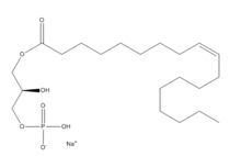
Lysophosphatidic Acid
Overall Structure
SHOC2
PP1C
MRAS
Key Ligand Interactions
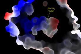
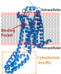
Figure 3: Electrostatic illustration of the amphipathic binding pocket of the LPA1 receptor. This binding pocket was revealed by cutting away the exterior or the protein. This binding pocket, located in the interior of the protein, has both polar and nonpolar regions. The blue and red coloration highlight the positively and negatively charged regions, respectively, and the white color shows the nonpolar region of the binding pocket.
SHOC2 and PP1C
SHOC2 and MRAS
PP1C and MRAS
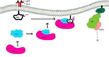
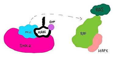
Signaling Pathway
Disease Relevance
Cancer
RASopathies
Future Studies
3D structures of lysophosphatidic acid receptor
4z34, 4z35, 4z36 - hLPA1 + antagonist - human
2lq4 – hLPA1 second extracellular loop – NMR
4p0c – hLPA2/NHERF2
5xsz – LPA6A (mutant) – zebra fish
References
- ↑ Hauseman ZJ, Fodor M, Dhembi A, Viscomi J, Egli D, Bleu M, Katz S, Park E, Jang DM, Porter KA, Meili F, Guo H, Kerr G, Molle S, Velez-Vega C, Beyer KS, Galli GG, Maira SM, Stams T, Clark K, Eck MJ, Tordella L, Thoma CR, King DA. Structure of the MRAS-SHOC2-PP1C phosphatase complex. Nature. 2022 Jul 13. pii: 10.1038/s41586-022-05086-1. doi:, 10.1038/s41586-022-05086-1. PMID:35830882 doi:http://dx.doi.org/10.1038/s41586-022-05086-1
- ↑ Kwon JJ, Hajian B, Bian Y, Young LC, Amor AJ, Fuller JR, Fraley CV, Sykes AM, So J, Pan J, Baker L, Lee SJ, Wheeler DB, Mayhew DL, Persky NS, Yang X, Root DE, Barsotti AM, Stamford AW, Perry CK, Burgin A, McCormick F, Lemke CT, Hahn WC, Aguirre AJ. Structure-function analysis of the SHOC2-MRAS-PP1C holophosphatase complex. Nature. 2022 Jul 13. pii: 10.1038/s41586-022-04928-2. doi:, 10.1038/s41586-022-04928-2. PMID:35831509 doi:http://dx.doi.org/10.1038/s41586-022-04928-2
- ↑ Lavoie H, Therrien M. Structural keys unlock RAS-MAPK cellular signalling pathway. Nature. 2022 Sep;609(7926):248-249. PMID:35970881 doi:10.1038/d41586-022-02189-7
- ↑ Liau NPD, Johnson MC, Izadi S, Gerosa L, Hammel M, Bruning JM, Wendorff TJ, Phung W, Hymowitz SG, Sudhamsu J. Structural basis for SHOC2 modulation of RAS signalling. Nature. 2022 Jun 29. pii: 10.1038/s41586-022-04838-3. doi:, 10.1038/s41586-022-04838-3. PMID:35768504 doi:http://dx.doi.org/10.1038/s41586-022-04838-3
Proteopedia Resources
Category:Lysophosphatidic acid binding
Category:Lysophosphatidic acid
Butler University Proteopedia Pages
See also:
</StructureSection>
Student Contributors
Madeline Gilbert Inaya Patel Rushda Hussein
