Sandbox Reserved 1771
From Proteopedia
| Line 9: | Line 9: | ||
===Antigen Binding Site=== | ===Antigen Binding Site=== | ||
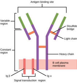
[[Image:B cell diagram.jpg| 250 px| thumb|right|'''Figure 1.''' Overview of the human B Cell Receptor and its structural components. Used with permission under Wikimedia Commons.]] | [[Image:B cell diagram.jpg| 250 px| thumb|right|'''Figure 1.''' Overview of the human B Cell Receptor and its structural components. Used with permission under Wikimedia Commons.]] | ||
| - | The binding of an antigen to the human B Cell receptor is identical to other common soluble antibodies (such as [https://en.wikipedia.org/wiki/Immunoglobulin_G IgG], [https://en.wikipedia.org/wiki/Immunoglobulin_A IgA], [https://en.wikipedia.org/wiki/Immunoglobulin_M IgM], [https://en.wikipedia.org/wiki/Immunoglobulin_E IgE], or [https://en.wikipedia.org/wiki/Immunoglobulin_D IgD]). The antibody portion of the B Cell Receptor is roughly "Y" shaped and consists of two identical <scene name='95/952701/Heavy_chains_highlight/2'>heavy</scene> and two identical <scene name='95/952701/Light_chains_highlight/3'>light</scene> chains creating two similar epitope or binding regions. Thus, two antigen molecules can bind independent of one another to produce a response. Within this structure, there are both constant and variable regions. The stem of the "Y" is a <scene name='95/952701/Constant_stem/1'>constant region</scene> ([https://en.wikipedia.org/wiki/Antibody#CDRs,_Fv,_Fab_and_Fc_Regions Fc]) composed of only heavy chain interactions. The two heavy chains then branch at a flexible <scene name='95/952701/Hinge/1'>hinge region</scene>. These interact individually with one light chain creating two [https://en.wikipedia.org/wiki/Antibody#CDRs,_Fv,_Fab_and_Fc_Regions Fab] fragments or branches of the "Y". Each <scene name='95/952701/Fab/1'>Fab fragment</scene> additionally contains a <scene name='95/952701/Fv_region/1'>variable region</scene> ([https://en.wikipedia.org/wiki/Antibody#CDRs,_Fv,_Fab_and_Fc_Regions Fv]) and a <scene name='95/952701/Fab_constant/1'>constant region</scene>. The variable region sits on top of the constant region and consists of hyper-variable loops which are random coils of amino acids that are unique to an antibody and exposed to allow specific recognition of an antigen. Furthermore, the light chains interact with the heavy chains via weak intermolecular forces and disulfide bridges. Therefore, binding to an antigen is processed through intermolecular interactions and is specific due to unique hyper variable loop sequences. | + | The binding of an antigen to the human B Cell receptor is identical to other common soluble antibodies (such as [https://en.wikipedia.org/wiki/Immunoglobulin_G IgG], [https://en.wikipedia.org/wiki/Immunoglobulin_A IgA], [https://en.wikipedia.org/wiki/Immunoglobulin_M IgM], [https://en.wikipedia.org/wiki/Immunoglobulin_E IgE], or [https://en.wikipedia.org/wiki/Immunoglobulin_D IgD]). The antibody portion of the B Cell Receptor is roughly "Y" shaped and consists of two identical <scene name='95/952701/Heavy_chains_highlight/2'>heavy</scene> and two identical <scene name='95/952701/Light_chains_highlight/3'>light</scene> chains creating two similar epitope or binding regions<Ref name="Janeway CA">Janeway CA Jr, Travers P, Walport M, et al. Immunobiology: The Immune System in Health and Disease. 5th edition. New York: Garland Science; 2001. </Ref>. Thus, two antigen molecules can bind independent of one another to produce a response. Within this structure, there are both constant and variable regions. The stem of the "Y" is a <scene name='95/952701/Constant_stem/1'>constant region</scene> ([https://en.wikipedia.org/wiki/Antibody#CDRs,_Fv,_Fab_and_Fc_Regions Fc]) composed of only heavy chain interactions<Ref name="Janeway CA">Janeway CA Jr, Travers P, Walport M, et al. Immunobiology: The Immune System in Health and Disease. 5th edition. New York: Garland Science; 2001. </Ref>. The two heavy chains then branch at a flexible <scene name='95/952701/Hinge/1'>hinge region</scene>. These interact individually with one light chain creating two [https://en.wikipedia.org/wiki/Antibody#CDRs,_Fv,_Fab_and_Fc_Regions Fab] fragments or branches of the "Y"<Ref name="Janeway CA">Janeway CA Jr, Travers P, Walport M, et al. Immunobiology: The Immune System in Health and Disease. 5th edition. New York: Garland Science; 2001. </Ref>. Each <scene name='95/952701/Fab/1'>Fab fragment</scene> additionally contains a <scene name='95/952701/Fv_region/1'>variable region</scene> ([https://en.wikipedia.org/wiki/Antibody#CDRs,_Fv,_Fab_and_Fc_Regions Fv]) and a <scene name='95/952701/Fab_constant/1'>constant region</scene><Ref name="Janeway CA">Janeway CA Jr, Travers P, Walport M, et al. Immunobiology: The Immune System in Health and Disease. 5th edition. New York: Garland Science; 2001. </Ref>. The variable region sits on top of the constant region and consists of hyper-variable loops which are random coils of amino acids that are unique to an antibody and exposed to allow specific recognition of an antigen<Ref name="Janeway CA">Janeway CA Jr, Travers P, Walport M, et al. Immunobiology: The Immune System in Health and Disease. 5th edition. New York: Garland Science; 2001. </Ref>. Furthermore, the light chains interact with the heavy chains via weak intermolecular forces and disulfide bridges<Ref name="Janeway CA">Janeway CA Jr, Travers P, Walport M, et al. Immunobiology: The Immune System in Health and Disease. 5th edition. New York: Garland Science; 2001. </Ref>. Therefore, binding to an antigen is processed through intermolecular interactions and is specific due to unique hyper variable loop sequences. |
===Fc and α/β Interactions=== | ===Fc and α/β Interactions=== | ||
Revision as of 21:58, 29 March 2023
| This Sandbox is Reserved from February 27 through August 31, 2023 for use in the course CH462 Biochemistry II taught by R. Jeremy Johnson at the Butler University, Indianapolis, USA. This reservation includes Sandbox Reserved 1765 through Sandbox Reserved 1795. |
To get started:
More help: Help:Editing |
Contents |
IgM B-cell Receptor
Introduction
B-cells play an important role of the human immune system and can be found circulating throughout the body. On the surface of B-cells, membrane bound B-cell receptors(BCRs) can be found[1]. These complex proteins are made up of membrane bound immunoglobulins (mIg). There are several different types of BCRs, namely IgG, IgA, IgM, IgE, or IgD. Each specific BCR has important functions for different diseases, but the IgM BCR in particular is most interesting. The BCR consists mainly of three domains: extracellular, transmembrane, and intracellular. While the extracellular region makes up most of the protein, perhaps the most interesting interactions can be found in the transmembrane domain. Unlike other BCRs, the IgM BCR has a specific heavy chain interaction with the α-β subunit of the protein[2]. The role of BCRs is to bind to foreign antigens and initiate the appropriate immune response.
Structure
| |||||||||||
Function
Once bound to an antigen, BCRs undergo a conformational change in the extracellular region. While the exact conformational change is still not known, preliminary studies suggest that there is separation of Fab fragments that opens the binding site within the BCR(ref). This initiates several signal transduction pathways, which are responsible for processing the antigen and initiating the appropriate immune responses. More specifically, the α-β subunit is connected to the phosphorylation of an immunoreceptor tyrosine-based activation motif(ITAM) upon binding. This in turn triggers the activation of kinases downstream that aid in the immune response. BCRs can be oligomeric prior to antigen binding, but once bound become an active monomer. [5].
Medical Relevancy
B-cell Formation
The formation of B-cells occurs in the bone marrow from hematopoietic stem cells[6]. Once formed, B-cell receptors are attached to B-cells through the aid of membrane-bound proteins in bone marrow cells. During this process, gene recombination occurs, which allows unique BCRs to become highly specific to different antigens.
Diseases
Therapeutics
References
- ↑ Robinson R. Distinct B cell receptor functions are determined by phosphorylation. PLoS Biol. 2006 Jul;4(7):e231. PMID:20076604 doi:10.1371/journal.pbio.0040231
- ↑ 2.0 2.1 Su Q, Chen M, Shi Y, Zhang X, Huang G, Huang B, Liu D, Liu Z, Shi Y. Cryo-EM structure of the human IgM B cell receptor. Science. 2022 Aug 19;377(6608):875-880. [doi: 10.1126/science.abo3923. Epub 2022 Aug 18. PMID: 35981043.]
- ↑ 3.0 3.1 3.2 3.3 3.4 3.5 Janeway CA Jr, Travers P, Walport M, et al. Immunobiology: The Immune System in Health and Disease. 5th edition. New York: Garland Science; 2001.
- ↑ 4.0 4.1 Tolar P, Pierce SK. Unveiling the B cell receptor structure. Science. 2022 Aug 19;377(6608):819-820. [doi: 10.1126/science.add8065. Epub 2022 Aug 18. PMID: 35981020.]
- ↑ ShenSichen Z, LiZhengpeng L, Liu W,(2019) Conformational change within the extracellular domain of B cell receptor in B cell activation upon antigen binding [eLife 8:e42271. https://doi.org/10.7554/eLife.42271]
- ↑ Althwaiqeb, S. Histology, B Cell Lymphocyte; StatPearls Publishing, 2023.

