We apologize for Proteopedia being slow to respond. For the past two years, a new implementation of Proteopedia has been being built. Soon, it will replace this 18-year old system. All existing content will be moved to the new system at a date that will be announced here.
Sandbox Reserved 1777
From Proteopedia
(Difference between revisions)
| Line 7: | Line 7: | ||
=Structure= | =Structure= | ||
==Overview== | ==Overview== | ||
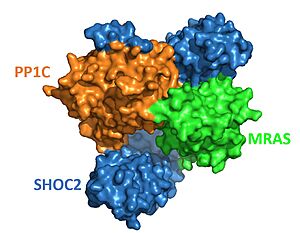
| - | [[Image:SHOC2-PP1C-MRAS Surface.JPG|300px|right|thumb|<font size="3.5"><div style="text-align: center;">'''Figure 1'''. Surface representation of SHOC2-PP1C-MRAS from PDB | + | [[Image:SHOC2-PP1C-MRAS Surface.JPG|300px|right|thumb|<font size="3.5"><div style="text-align: center;">'''Figure 1'''. Surface representation of SHOC2-PP1C-MRAS from PDB 7pui. Blue is SHOC2, orange is PP1C and green is MRAS. </div></font>]] |
| - | The enzyme requires 3 domains, | + | The enzyme requires 3 domains, SHOC2(blue), PP1C(orange), and MRAS(green), to form the active enzyme (SMP Complex), also known as a holoenzyme (Figure 1)<Ref name='Hauseman'>Hauseman, Z.J., Fodor, M., Dhembi, A. et al. Structure of the MRAS–SHOC2–PP1C phosphatase complex. Nature 609, 416–423 (2022). doi: 10.1038/s41586-022-05086-1. [https://doi.org/10.1038/s41586-022-05086-1. DOI:10.1038/s41586-022-05086-1]. </Ref>. The SMP complex was determined via cryo-electron microscopy as well as x-ray diffraction. These studies found that PP1C and MRAS occupy the concave surface of SHOC2, leaving the catalytic site of PP1C and the substrate binding cleft in MRAS exposed. |
==SHOC2== | ==SHOC2== | ||
| Line 18: | Line 18: | ||
==MRAS== | ==MRAS== | ||
| - | <scene name='95/952705/Mras_structure/4'>MRAS</scene> is a GTPase protein and is | + | <scene name='95/952705/Mras_structure/4'>MRAS</scene> is a GTPase protein and is anchored in the cell membrane. When MRAS binds GTP, it becomes active and triggers the assembly of the active holoenzyme<ref name="Hauseman" />. MRAS is a close relative of the RAS protein and therefore shares most of it's regulatory and effector interactions<Ref name= 'Young'>Young, L., Rodriguez-Viciana, P. MRAS: A Close but Understudied Member of the RAS Family. Cold Spring Harbor Perspectives in Medicine (2018). doi: 10.1101/cshperspect.a033621. [https://perspectivesinmedicine.cshlp.org/content/8/12/a033621.full.pdf+html. DOI: 0.1101/cshperspect.a033621]. </Ref>. A unique function of MRAS is it's ability to form a complex with SHOC2 and PP1, allowing it to have phosphatase activity. |
===Switch I and II=== | ===Switch I and II=== | ||
| - | Switch I and II are located in MRAS. The switches determine whether MRAS can bind to SHOC2-PP1C. The switches have to go through a conformational change to allow binding of SHOC2-PP1C to MRAS. This conformational change is caused by GTP replacing GDP. Once GTP is added MRAS shifts and binds with SHOC2. When GDP is bound to MRAS switch II is moved outward which causes a steric clash with SHOC2 <scene name='95/952705/Switch_i_and_ii_with_gdp/8'>(Switch I and II with GDP)</scene>. When GTP is bound, switch II can form various hydrophobic interactions with SHOC2<Ref name='Bonsor'>Daniel A. Bonsor, Patrick Alexander, Kelly Snead, Nicole Hartig, Matthew Drew, Simon Messing, Lorenzo I. Finci, Dwight V. Nissley, Frank McCormick, Dominic Esposito, Pablo Rodrigiguez-Viciana, Andrew G. Stephen, Dhirendra K. Simanshu. Structure of the SHOC2–MRAS–PP1C complex provides insights into RAF activation and Noonan syndrome. bioRxiv. 2022.05.10.491335. doi: 10.1101/2022.05.10.491335. [https://doi.org/10.1101/2022.05.10.491335. DOI:10.1101/2022.05.10.491335]. </Ref> <scene name='95/952705/Switch_i_and_ii_with_gtp/5'>(Switch I and II with GTP)</scene>. Interactions are strengthened with hydrogen bonding and pi stacking. When MRAS is bound to SHOC2-PP1C, switch I has an important role in making interactions with PP1C. | + | Switch I and II are located in MRAS. The switches determine whether MRAS can bind to SHOC2-PP1C. The switches have to go through a conformational change to allow binding of SHOC2-PP1C to MRAS. This conformational change is caused by GTP replacing GDP. Once GTP is added MRAS shifts and binds with SHOC2. When GDP is bound to MRAS, switch II is moved outward which causes a steric clash with SHOC2 <scene name='95/952705/Switch_i_and_ii_with_gdp/8'>(Switch I and II with GDP)</scene>. When GTP is bound, switch II can form various hydrophobic interactions with SHOC2<Ref name='Bonsor'>Daniel A. Bonsor, Patrick Alexander, Kelly Snead, Nicole Hartig, Matthew Drew, Simon Messing, Lorenzo I. Finci, Dwight V. Nissley, Frank McCormick, Dominic Esposito, Pablo Rodrigiguez-Viciana, Andrew G. Stephen, Dhirendra K. Simanshu. Structure of the SHOC2–MRAS–PP1C complex provides insights into RAF activation and Noonan syndrome. bioRxiv. 2022.05.10.491335. doi: 10.1101/2022.05.10.491335. [https://doi.org/10.1101/2022.05.10.491335. DOI:10.1101/2022.05.10.491335]. </Ref> <scene name='95/952705/Switch_i_and_ii_with_gtp/5'>(Switch I and II with GTP)</scene>. Interactions are strengthened with hydrogen bonding and pi stacking. When MRAS is bound to SHOC2-PP1C, switch I has an important role in making interactions with PP1C. |
==Special Interactions== | ==Special Interactions== | ||
| - | MRAS binds to SHOC2 exclusively through this concave region <Ref name='Kwan'>Kwon, J.J., Hajian, B., Bian, Y. et al. Structure–function analysis of the SHOC2–MRAS–PP1C holophosphatase complex. Nature 609, 408–415 (2022).doi: 10.1038/s41586-022-04928-2. [https://doi.org/10.1038/s41586-022-04928-2. DOI:10.1038/s41586-022-04928-2] </Ref>, primarily by the descending loop and strands of each LRR domains 2-10. This reaction is stabilized through <scene name='95/952705/Shoc2_mras_interaction/2'>hydrogen bonding</scene> between residues of each subunit. <scene name='95/952705/Shoc2_pp1c_interaction/1'>PP1C binds</scene> with the ascending loops of the SHOC2 LRR regions, and is further engaged through the N-terminal region containing the <scene name='95/952705/Shoc2_rvxf/3'>PVxF motif</scene> <Ref name= 'Jajian'>Kwon, J., Jajian, B., Bian, Y. et al. Comprehensive structure-function evaluation of the SHOC2 holophosphatase reveals disease mechanisms and therapeutic opportunities. In: Proceedings of the American Association for Cancer Research Annual Meeting 2022. [https://aacrjournals.org/cancerres/article/82/12_Supplement/LB029/699443. DOI: 10.1158/1538-7445.AM2022-LB029]. </Ref>. The initial forming of the complex begins with SHOC2 | + | MRAS binds to SHOC2 exclusively through this concave region <Ref name='Kwan'>Kwon, J.J., Hajian, B., Bian, Y. et al. Structure–function analysis of the SHOC2–MRAS–PP1C holophosphatase complex. Nature 609, 408–415 (2022).doi: 10.1038/s41586-022-04928-2. [https://doi.org/10.1038/s41586-022-04928-2. DOI:10.1038/s41586-022-04928-2] </Ref>, primarily by the descending loop and strands of each LRR domains 2-10. This reaction is stabilized through <scene name='95/952705/Shoc2_mras_interaction/2'>hydrogen bonding</scene> between residues of each subunit. <scene name='95/952705/Shoc2_pp1c_interaction/1'>PP1C binds</scene> with the ascending loops of the SHOC2 LRR regions, and is further engaged through the N-terminal region containing the <scene name='95/952705/Shoc2_rvxf/3'>PVxF motif</scene> <Ref name= 'Jajian'>Kwon, J., Jajian, B., Bian, Y. et al. Comprehensive structure-function evaluation of the SHOC2 holophosphatase reveals disease mechanisms and therapeutic opportunities. In: Proceedings of the American Association for Cancer Research Annual Meeting 2022. [https://aacrjournals.org/cancerres/article/82/12_Supplement/LB029/699443. DOI: 10.1158/1538-7445.AM2022-LB029]. </Ref>. The initial forming of the complex begins with SHOC2-PP1C engagement, then is completed and stabilized by the GTP-loaded MRAS binding <Ref name="Jajian" />. Once associated with SHOC2, <scene name='95/952705/Mras_pp1c_interaction/2'>MRAS binds to PP1C</scene> and guides the holoenzyme complex to the cell membrane to begin signaling<ref name="Kwan" />. |
=Mechanism= | =Mechanism= | ||
[[Image: Image 4-3-23 at 10.02 AM.jpeg|300px|right|thumb|<font size="3.5"><div style="text-align: center;">Schematic representation of SHOC2-PP1C-MRAS pathway after all domains are bound together. </div></font>]] | [[Image: Image 4-3-23 at 10.02 AM.jpeg|300px|right|thumb|<font size="3.5"><div style="text-align: center;">Schematic representation of SHOC2-PP1C-MRAS pathway after all domains are bound together. </div></font>]] | ||
| - | Once the SMP complex comes together, it plays a key role in regulating the activation of the RAF and MAPK pathway. To do so, PP1C, enhanced through the interactions with SHOC2 and MRAS, | + | Once the SMP complex comes together, it plays a key role in regulating the activation of the RAF and MAPK pathway. To do so, PP1C, enhanced through the interactions with SHOC2 and MRAS, dephosphorylates a specific phosphoserine on RAF kinases. Doing so regulates cell growth, survival, proliferation, and differentiation <Ref name='Lavoie'>Lavoie, H., Therrien, M. Structural keys unlock RAS–MAPK cellular signalling pathway. Nature 609, 248-249 (2022). doi: 10.1038/d41586-022-02189-7. [https://doi.org/10.1038/d41586-022-02189-7. DOI:10.1038/d41586-022-02189-7]. </Ref>. |
==Active Site== | ==Active Site== | ||
Revision as of 19:05, 3 April 2023
| This Sandbox is Reserved from February 27 through August 31, 2023 for use in the course CH462 Biochemistry II taught by R. Jeremy Johnson at the Butler University, Indianapolis, USA. This reservation includes Sandbox Reserved 1765 through Sandbox Reserved 1795. |
To get started:
More help: Help:Editing |
| |||||||||||
References
- ↑ 1.0 1.1 Bernal Astrain G, Nikolova M, Smith MJ. Functional diversity in the RAS subfamily of small GTPases. Biochem Soc Trans. 2022 Apr 29;50(2):921-933. doi: 10.1042/BST20211166. DOI:10.1042/BST20211166
- ↑ Molina JR, Adjei AA. The Ras/Raf/MAPK pathway. J Thorac Oncol. 2006 Jan;1(1):7-9. DOI:10.1016/S1556-0864(15)31506-9.
- ↑ Li, L., Zhao, G. D., Shi, Z. et. al.The Ras/Raf/MEK/ERK signaling pathway and its role in the occurrence and development of HCC. Oncology letters, 12(5), 3045–3050. DOI:10.3892/ol.2016.5110.
- ↑ 4.0 4.1 Hauseman, Z.J., Fodor, M., Dhembi, A. et al. Structure of the MRAS–SHOC2–PP1C phosphatase complex. Nature 609, 416–423 (2022). doi: 10.1038/s41586-022-05086-1. DOI:10.1038/s41586-022-05086-1.
- ↑ Young, L., Rodriguez-Viciana, P. MRAS: A Close but Understudied Member of the RAS Family. Cold Spring Harbor Perspectives in Medicine (2018). doi: 10.1101/cshperspect.a033621. DOI: 0.1101/cshperspect.a033621.
- ↑ Daniel A. Bonsor, Patrick Alexander, Kelly Snead, Nicole Hartig, Matthew Drew, Simon Messing, Lorenzo I. Finci, Dwight V. Nissley, Frank McCormick, Dominic Esposito, Pablo Rodrigiguez-Viciana, Andrew G. Stephen, Dhirendra K. Simanshu. Structure of the SHOC2–MRAS–PP1C complex provides insights into RAF activation and Noonan syndrome. bioRxiv. 2022.05.10.491335. doi: 10.1101/2022.05.10.491335. DOI:10.1101/2022.05.10.491335.
- ↑ 7.0 7.1 Kwon, J.J., Hajian, B., Bian, Y. et al. Structure–function analysis of the SHOC2–MRAS–PP1C holophosphatase complex. Nature 609, 408–415 (2022).doi: 10.1038/s41586-022-04928-2. DOI:10.1038/s41586-022-04928-2
- ↑ 8.0 8.1 8.2 Kwon, J., Jajian, B., Bian, Y. et al. Comprehensive structure-function evaluation of the SHOC2 holophosphatase reveals disease mechanisms and therapeutic opportunities. In: Proceedings of the American Association for Cancer Research Annual Meeting 2022. DOI: 10.1158/1538-7445.AM2022-LB029.
- ↑ 9.0 9.1 Lavoie, H., Therrien, M. Structural keys unlock RAS–MAPK cellular signalling pathway. Nature 609, 248-249 (2022). doi: 10.1038/d41586-022-02189-7. DOI:10.1038/d41586-022-02189-7.
- ↑ Liau NPD, Johnson MC, Izadi S, Gerosa L, Hammel M, Bruning JM, Wendorff TJ, Phung W, Hymowitz SG, Sudhamsu J. Structural basis for SHOC2 modulation of RAS signalling. Nature. 2022 Jun 29. pii: 10.1038/s41586-022-04838-3. doi:, 10.1038/s41586-022-04838-3. PMID:35768504 doi:http://dx.doi.org/10.1038/s41586-022-04838-3


