We apologize for Proteopedia being slow to respond. For the past two years, a new implementation of Proteopedia has been being built. Soon, it will replace this 18-year old system. All existing content will be moved to the new system at a date that will be announced here.
Image:Mechanism.png
From Proteopedia
(Difference between revisions)

No higher resolution available.
Mechanism.png (773 × 600 pixel, file size: 68 KB, MIME type: image/png)
(uploaded a new version of "Image:Mechanism.png") |
(uploaded a new version of "Image:Mechanism.png") |
Revision as of 19:24, 25 March 2025
Proteopedia Page Contributors and Editors (what is this?)
Joseph Gareis, Rushda Hussein, Hayden Vissing, Charles Short, Hanan Busaileh, Francis Ayombil, Jamie C. Gladfelder, Zhichang Yang, Cory Tiedeman
File history
Click on a date/time to view the file as it appeared at that time.
| Date/Time | User | Dimensions | File size | Comment | |
|---|---|---|---|---|---|
| (current) | 19:35, 25 March 2025 | Hayden Vissing (Talk | contribs) | 773×600 | 68 KB | |
| 19:32, 25 March 2025 | Hayden Vissing (Talk | contribs) | 1308×1015 | 137 KB | ||
| 19:30, 25 March 2025 | Hayden Vissing (Talk | contribs) | 1308×1015 | 137 KB | ||
| 19:27, 25 March 2025 | Hayden Vissing (Talk | contribs) | 1308×1015 | 137 KB | ||
| 19:24, 25 March 2025 | Hayden Vissing (Talk | contribs) | 1308×1015 | 137 KB | ||
| 18:57, 6 April 2023 | Rushda Hussein (Talk | contribs) | 2182×1538 | 1.14 MB | ||
| 18:55, 6 April 2023 | Rushda Hussein (Talk | contribs) | 2182×1538 | 1.14 MB | ||
| 03:29, 18 April 2022 | Joseph Gareis (Talk | contribs) | 1478×762 | 294 KB | ||
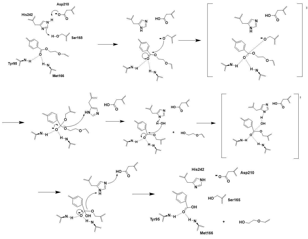
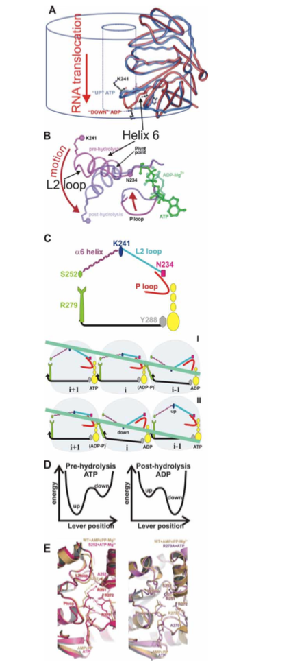
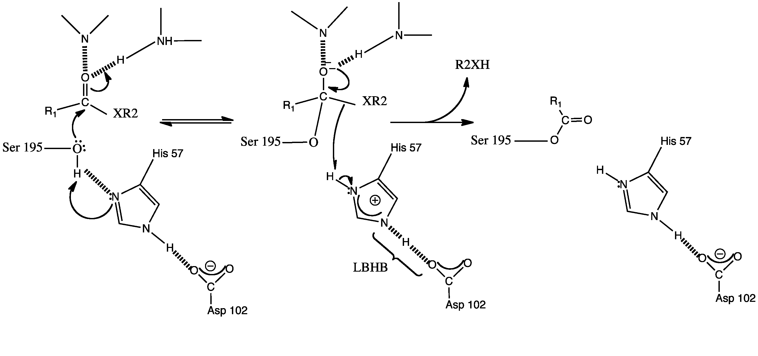
| 13:37, 17 December 2015 | Student (Talk | contribs) | 428×968 | 319 KB | (A–B) Loop L2 movement in the central channel (C) Mechanism of sequential hydrolysis of ATP (yellow) and RNA (light green) translocation for three subunits viewed from within the central | |
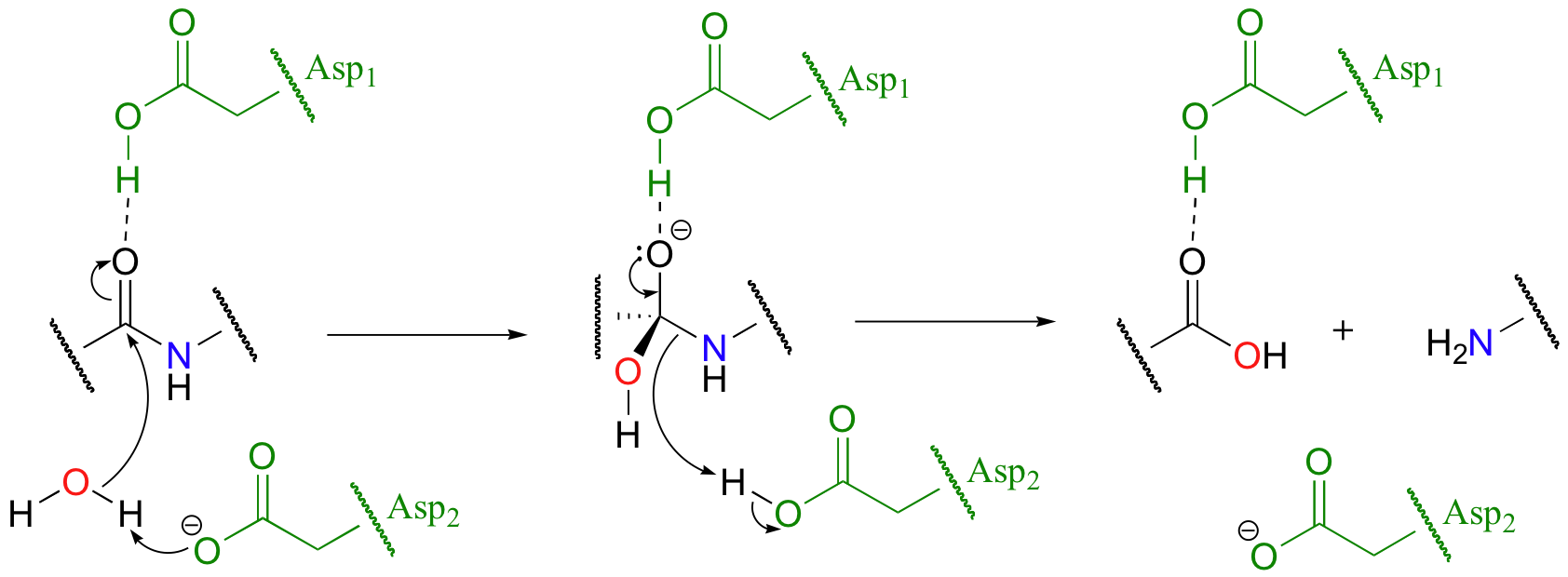
| 00:11, 27 November 2012 | Charles Short (Talk | contribs) | 1684×620 | 162 KB | mechanism | |
| 21:07, 25 November 2012 | Hanan Busaileh (Talk | contribs) | 647×260 | 51 KB | ||
| 21:07, 25 November 2012 | Hanan Busaileh (Talk | contribs) | 647×260 | 51 KB | ||
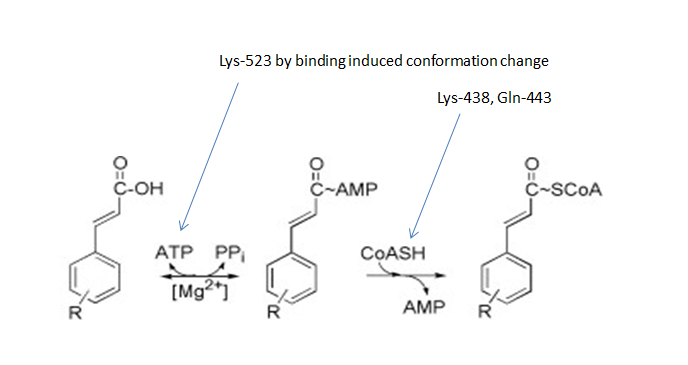
| 19:22, 19 November 2012 | Zhichang Yang (Talk | contribs) | 688×365 | 48 KB | ||
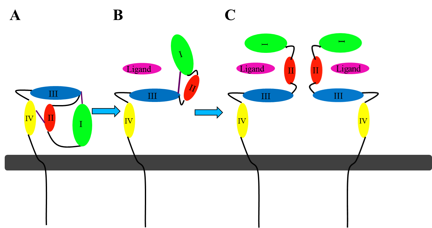
| 23:46, 6 November 2012 | Jamie C. Gladfelder (Talk | contribs) | 1378×729 | 55 KB | This picture illustrates the mechanism in which EGFR, HER3, and HER4 change conformation in order to dimerize and activate further cell signaling. A) Sub-domain I (green) forms an interaction (purple line) with sub-domain III (blue). Sub-domain II (red) f | |
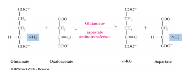
| 23:21, 14 April 2012 | Francis Ayombil (Talk | contribs) | 2876×1354 | 124 KB | mech 1 | |
| 05:43, 1 March 2010 | Cory Tiedeman (Talk | contribs) | 978×379 | 59 KB |
- Edit this file using an external application
See the setup instructions for more information.
Links
The following pages link to this file:
