This old version of Proteopedia is provided for student assignments while the new version is undergoing repairs. Content and edits done in this old version of Proteopedia after March 1, 2026 will eventually be lost when it is retired in about June of 2026.
Apply for new accounts at the new Proteopedia. Your logins will work in both the old and new versions.
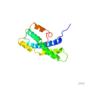
Lysin
From Proteopedia
(Difference between revisions)
| Line 8: | Line 8: | ||
== 3D structure of lysin == | == 3D structure of lysin == | ||
| + | |||
| + | [[Lysin 3D structures]] | ||
| + | |||
Updated on {{REVISIONDAY2}}-{{MONTHNAME|{{REVISIONMONTH}}}}-{{REVISIONYEAR}} | Updated on {{REVISIONDAY2}}-{{MONTHNAME|{{REVISIONMONTH}}}}-{{REVISIONYEAR}} | ||
Revision as of 10:32, 18 June 2023
| |||||||||||
References
- ↑ Shaw A, McRee DE, Vacquier VD, Stout CD. The crystal structure of lysin, a fertilization protein. Science. 1993 Dec 17;262(5141):1864-7. PMID:8266073
Proteopedia Page Contributors and Editors (what is this?)
Michal Harel, Carmit Ginesin, Alexander Berchansky, Joel L. Sussman, Jaime Prilusky
