Introduction
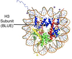
Figure 1: The crystal structure of a histone core octamer (each subunit in a different color) bound to DNA. PDB:5y0c
, human lysine-specific demethylase 1, is an enzyme that affects the ability of DNA to associate with histone proteins. Histone proteins contain residues that give them an overall positive charge and allow them to act as spools upon which negatively charged DNA can wrap around for storage in the nucleus of a cell (Figure 1). When DNA is tightly condensed it forms into nucleosomes which consist of eight histone core proteins (2 H2A, 2 H2B, 2 H3, 2 H4) with DNA tightly coiled around them. This tightly coiled DNA is known as heterochromatin, which is inaccessible to transcription factors and RNA polymerase. This can be reversed by post-translational modifications to the histone protein structure that cause the DNA to relax and form euchromatin, which allows for gene expression. One key histone modification is the methylation and subsequent demethylation of lysine residues. Before 2004, it was believed that methylation of histone tails was stable and irreversible. In 2004, it was discovered that histone tails can also be demethylated by demethylase enzymes such as LSD-1.[1]
There are two main classes of demethylases, and they are categorized by their cofactors and reaction mechanisms. The KDM1 family of demethylases employs a flavin adenine dinucleotide (FAD) cofactor to catalyze the demethylation reaction. The other class of demethylases consists of five families (KDM2-6) and use an Fe2+ ion cofactor and α-ketoglutarate as a cosubstrate to catalyze the reaction. Although the cofactors used are different, both classes operate by hydroxylating the target methyl group. LSD-1 is in the KDM1 family of histone demethylases that uses FAD as a cofactor. LSD-1 specifically demethylates mono- or di-methylated lysine substrates at Lys4 or Lys9 in the tail of histone H3.[2] Demethylation of these lysine residues is commonly associated with transcriptional activation, but it also has the ability to silence genes depending on the residue being demethylated, the cofactors present, and the environment in which the demethylation occurs.
Structure
The human lysine-specific demethylase-1 structure (PDB: 2H94) was determined to a resolution of 2.9 Å. The crystals were grown without the first 165 residues due to their proposed lack of structure and protease susceptibility. Additionally, two unstructured loops (residues 467–474 and 784–792) remained unresolved in the final refined structure.[3]
Tower Domain
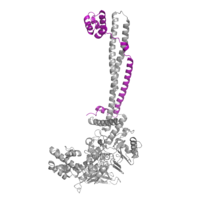
Figure 2: CoRest complex (purple) bound to LSD-1 (PDB: 2IW5) at the Tower domain.
The is a 100 residue protrusion off of the main protein body that is caused by an insertion into the sequence of the oxidase domain. It is comprised of a coiled-coil of two α-helices. The longer helix, TαA, is an LSD-1 specific element that has not been found in other oxidase proteins.[3] The shorter helix, TαB, connects directly to helix αD of the oxidase domain through a conserved connector loop. The base of the tower domain forms a direct connection to the oxidase domain and plays a crucial role in the shape and catalytic activity of the active site. Removing the tower domain via a mutation resulted in a decrease in catalytic efficiency.[3] The orientation of the tower domain, through the is responsible for positioning of Phe538 in the catalytic chamber, where it is proposed to orient the substrate lysine through hydrophobic interactions. In addition, the TαB-αD connector positions the αD in the correct manner to provide a hydrogen bond to Tyr761. Tyr761 is positioned in the catalytic chamber next to the FAD cofactor and aids in the binding of the lysine substrate.[3] The tower domain has also been found to interact with other proteins and complexes, such as CoREST (Figure 2), as a switch to allosterically regulate the catalytic activity of the protein.[4]
SWIRM Domain
The , named after the SWI3, RSC8, and MOIRA proteins from which it was first discovered,[5] is seen in numerous enzymes that participate in histone binding and chromatin modification. The SWIRM domain of LSD-1 is 94 residues and is comprised of an α-helix bundle. Two helix-turn-helix motifs, , are separated by a longer helix, αC. The SWIRM domain is associated with the oxidase domain via hydrophobic Van der Waals interactions between a set of : the αA, αB, and αE motifs in the SWIRM domain and the αA, αB, αM motifs in the oxidase domain. The residues that create this hydrophobic interface (which spans nearly 1680 Ų) are nearly invariant among histone-modifying proteins. The hydrophobic interface between the oxidase and SWIRM domains creates a or tunnel that is also present in other chromatin modifying enzymes. Typically in other KMD enzymes, the cleft is responsible for binding to histone tail through the presence of positively charged residues. However, the SWIRM domain in LSD-1 is unique because the cleft that is formed by the hydrophobic SWIRM-oxidase interactions lacks the positively charged residues common in other enzymes.[3]
Oxidase Domain
The houses the catalytic site of LSD-1. This domain non-covalently binds the FAD cofactor and the substrate lysine on the H3 histone tail.[3] The FAD binding cavity is quite large (15 Å deep and around 25 Å wide) in relation to other oxidases that utilize FAD as a cofactor (Figure 3, left panel).[3] In comparison, polyamine oxidase, another FAD-dependent oxidase, has a catalytic chamber roughly 30 Å long but only a few angstroms wide.[6] The relatively large size of the LSD-1 active site cavity is to accommodate the first 15 residues of the histone H3 substrate (Figure 3, right panel).
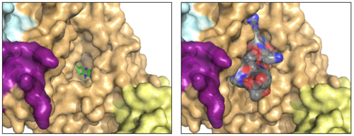
Figure 3: The FAD binding cavity in the oxidase domain of LSD-1 (left) and in the presence of histone H3-peptide (right). The swirm domain is yellow, CoREST is purple, the oxidase domain is orange, and the tower domain is light blue. The FAD is shown as green sticks and the H3-peptide is gray. PDB: 2V1D
FAD Cofactor
Within the active site cavity, there are (Val317, Gly330, Ala331, Met332, Val333, Phe538, Leu659, Asn660, Lys661, Trp695, Ser749, Ser760 and Tyr761) that facilitate the interaction between the FAD cofactor and the substrate lysine.[3] This pocket binds and positions the substrate lysine so that it is exposed to the FAD. During catalysis, the FAD is reduced and becomes anionic. To stabilize this negatively charged intermediate form of the cofactor, in most FAD-dependent oxidases a positively charged residue is found nearby. In LSD-1, is present in the catalytic pocket of the active site to help stabilize the negative charge that develops on the FAD as it is reduced.[3] The adenine-dinucleotide moiety of FAD is bound by a β-α-β Rossmann fold that is of the glycine rich consensus sequence Gly-Ser-Gly-Val-Ser-Gly spanning . As is typical of a Rossmann fold, this sequence is in the loop connecting the first β-strand (β1) with the α-helix.
Substrate Binding
A of the LSD-1/CoREST complex was determined bound to a 16-residue histone H3-peptide substrate.[7] In the substrate, the Lys4 target was mutated to methionine (Met4). The was revealed to lie between the flavin ring of the FAD cofactor and helices αC and αD of the oxidase domain. The substrate makes one helical turn at its N terminus and makes extensive intra- and intermolecular interactions with itself and the oxidase domain using two arginine residues, and
. The of the H3-peptide is nearly buried from solvent by a short (one turn), acidic helix spanning residues 555-559 of the oxidase domain.
Enzymatic Mechanism
The mechanism of lysine demethylation catalyzed by LSD-1 employs FAD as an oxidizing agent to oxidize the methyl carbon of the lysine substrate (Figure 4). The FAD cofactor, positioned close to the substrate lysine in the active site, initiates a two-electron transfer in the form of a hydride between the substrate methyl-lysine and the isoallozaxine ring of the FAD. The cofactor becomes anionic and the resonance form is stabilized by the positively charged positioned in the catalytic pocket of the active site. The lysine subatrate forms an iminium cation that is hydrolyzed into the hemiaminal intermediate, potentially using water and general base catalysis of a nearby residue. The hemiaminal intermediate readily decomposes to formaldehyde and the demethylated lysine.[3]
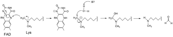
Figure 4: Hydride transfer mechanism catalyzed by LSD-1 with a dimethyl-lysine substrate.
Inhibition by Tri-Methylated Lysine
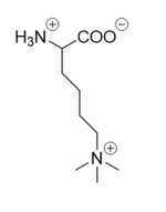
Figure 5: Tri-methylated lysine.
The proposed LSD-1 mechanism is supported by the fact that tri-methylated lysine substrates (Figure 5) competitively inhibit the enzyme. A substrate lysine that is tri-methylated binds to the active site but does not undergo catalysis. The resolved enzyme structure reveals the inhibition is not steric as the active site is large enough to accommodate tri-methylated lysines, but rather is chemical in nature. Tri-methylated lysines do not have a free lone pair to form an iminium cation as is necessitated by the proposed mechanism, resulting in chemical inhibition of LSD-1.[3] Thus, the mechanism of LSD-1 contributes to its specificity for mono- or di-methylated lysine substrates.
Medical Implications
Diabetes
Gluconeogenesis is a process that results in the production of glucose from non-carbohydrate compounds. Glucose 6-phosphatase (G6Pase) and fructose 1,6-bisphosphatase 1 (FBP1) are critical enzymes in the gluconeogenesis pathway. LSD-1, although it can have both activating and inhibiting effects depending on external conditions, is proposed to have inhibiting effects on the transcription of both G6Pase and FBP1.[8] Under healthy conditions, LSD-1 inhibits the transcription of these enzymes in order to regulate the blood glucose levels in the body. It was found that decreased amounts of LSD-1 can induce hyperglycemia that contributes to the onset of diabetes.[8]
Cancer
Research has found that LSD-1 is over-expressed in many tumorous cancers. The proposed mechanism behind the carcinogenic role of LSD-1 focuses on the known tumor-suppressor gene, p53. The p53 protein acts as a transcription factor that activates the expression of genes the control cellular proliferation and because of this, p53 is known as the "guardian of the genome."[9][10] LSD-1 has been found to remove a methyl group from the di-methylated Lys370 on p53.[11] Similar to the proposed role of LSD-1 in diabetes, its demethylation of p53 is inhibitory and prevents its binding to DNA.[11] This inactivation of p53 is thought to prevent anti-proliferative operations in the cell and contribute to the development of multiple types of cancers.
References
- ↑ Shi Y, Lan F, Matson C, Mulligan P, Whetstine JR, Cole PA, Casero RA, Shi Y. Histone demethylation mediated by the nuclear amine oxidase homolog LSD1. Cell. 2004 Dec 29;119(7):941-53. PMID:15620353 doi:http://dx.doi.org/10.1016/j.cell.2004.12.012
- ↑ Forneris F, Binda C, Vanoni MA, Mattevi A, Battaglioli E. Histone demethylation catalysed by LSD1 is a flavin-dependent oxidative process. FEBS Lett. 2005 Apr 11;579(10):2203-7. doi: 10.1016/j.febslet.2005.03.015. PMID:15811342 doi:http://dx.doi.org/10.1016/j.febslet.2005.03.015
- ↑ 3.00 3.01 3.02 3.03 3.04 3.05 3.06 3.07 3.08 3.09 3.10 Stavropoulos P, Blobel G, Hoelz A. Crystal structure and mechanism of human lysine-specific demethylase-1. Nat Struct Mol Biol. 2006 Jul;13(7):626-32. Epub 2006 Jun 25. PMID:16799558 doi:10.1038/nsmb1113
- ↑ Yang M, Gocke CB, Luo X, Borek D, Tomchick DR, Machius M, Otwinowski Z, Yu H. Structural basis for CoREST-dependent demethylation of nucleosomes by the human LSD1 histone demethylase. Mol Cell. 2006 Aug 4;23(3):377-87. PMID:16885027 doi:http://dx.doi.org/10.1016/j.molcel.2006.07.012
- ↑ Da G, Lenkart J, Zhao K, Shiekhattar R, Cairns BR, Marmorstein R. Structure and function of the SWIRM domain, a conserved protein module found in chromatin regulatory complexes. Proc Natl Acad Sci U S A. 2006 Feb 14;103(7):2057-62. Epub 2006 Feb 3. PMID:16461455
- ↑ Binda C, Angelini R, Federico R, Ascenzi P, Mattevi A. Structural bases for inhibitor binding and catalysis in polyamine oxidase. Biochemistry. 2001 Mar 6;40(9):2766-76. PMID:11258887
- ↑ Forneris F, Binda C, Adamo A, Battaglioli E, Mattevi A. Structural basis of LSD1-CoREST selectivity in histone H3 recognition. J Biol Chem. 2007 Jul 13;282(28):20070-4. Epub 2007 May 30. PMID:17537733 doi:10.1074/jbc.C700100200
- ↑ 8.0 8.1 Pan D, Mao C, Wang YX. Suppression of gluconeogenic gene expression by LSD1-mediated histone demethylation. PLoS One. 2013 Jun 5;8(6):e66294. doi: 10.1371/journal.pone.0066294. Print 2013. PMID:23755305 doi:http://dx.doi.org/10.1371/journal.pone.0066294
- ↑ Lane DP. Cancer. p53, guardian of the genome. Nature. 1992 Jul 2;358(6381):15-6. doi: 10.1038/358015a0. PMID:1614522 doi:http://dx.doi.org/10.1038/358015a0
- ↑ Toufektchan E, Toledo F. The Guardian of the Genome Revisited: p53 Downregulates Genes Required for Telomere Maintenance, DNA Repair, and Centromere Structure. Cancers (Basel). 2018 May 6;10(5). pii: cancers10050135. doi:, 10.3390/cancers10050135. PMID:29734785 doi:http://dx.doi.org/10.3390/cancers10050135
- ↑ 11.0 11.1 Jin L, Hanigan CL, Wu Y, Wang W, Park BH, Woster PM, Casero RA. Loss of LSD1 (lysine-specific demethylase 1) suppresses growth and alters gene expression of human colon cancer cells in a p53- and DNMT1(DNA methyltransferase 1)-independent manner. Biochem J. 2013 Jan 15;449(2):459-68. doi: 10.1042/BJ20121360. PMID:23072722 doi:http://dx.doi.org/10.1042/BJ20121360


