We apologize for Proteopedia being slow to respond. For the past two years, a new implementation of Proteopedia has been being built. Soon, it will replace this 18-year old system. All existing content will be moved to the new system at a date that will be announced here.
Sandbox Reserved 1849
From Proteopedia
(Difference between revisions)
| Line 6: | Line 6: | ||
===What are Minibinders?=== | ===What are Minibinders?=== | ||
These mini proteins target the interaction between ACE2 and COVID-19 spike protein <ref name="Longxing">PMID:32907861</ref>. The minibinders are small proteins carefully designed to bind to the COVID-19 spike protein with a greater affinity than ACE2 <ref name="Longxing">PMID:32907861</ref>. These minibinders were able to reduce the viral burden of SARS-CoV-2 in mice <ref name="Case">PMID:34192518</ref>. These proteins were de novo (from scratch) designs to mimic the ACE2 helix, but have a lower dissociation constant, yielding a greater affinity for the spike protein <ref name="Longxing">PMID:32907861</ref>. The binding region between <scene name='10/1075251/Ace2_and_rbd/2'>spike protein and ACE2</scene> can give a better explanation as to how these proteins were designed. | These mini proteins target the interaction between ACE2 and COVID-19 spike protein <ref name="Longxing">PMID:32907861</ref>. The minibinders are small proteins carefully designed to bind to the COVID-19 spike protein with a greater affinity than ACE2 <ref name="Longxing">PMID:32907861</ref>. These minibinders were able to reduce the viral burden of SARS-CoV-2 in mice <ref name="Case">PMID:34192518</ref>. These proteins were de novo (from scratch) designs to mimic the ACE2 helix, but have a lower dissociation constant, yielding a greater affinity for the spike protein <ref name="Longxing">PMID:32907861</ref>. The binding region between <scene name='10/1075251/Ace2_and_rbd/2'>spike protein and ACE2</scene> can give a better explanation as to how these proteins were designed. | ||
| - | [[Image:Untitled presentation.jpg]] | + | [[Image:Untitled presentation.jpg|100 px|right|thumb|Figure 1. Image of the individual helices of AHB2, LCB1, and LCB3, respectively.]] |
===COVID-19 Disease Pathway=== | ===COVID-19 Disease Pathway=== | ||
Understanding the pathway of the COVID-19 virus is essential to understanding the mechanism in which the virus’ surface proteins attach to the mini binders. The COVID-19 virus has spike proteins on its surface that bind to the host cell receptor, known as ACE2, and this allows the virus to remain anchored to the host for viral entry <ref name="Sang">PMID:36499120</ref>. When the spike protein binds to the receptor, ACE2 for example, the cell membrane-associated protease, protease serine 2 TMPRSS2 promotes viral entry by activating the spike protein <ref name="Huang">PMID:32747721</ref>. The activated spike protein is able to cleave itself into S1 and S2 subunits <ref name="Huang">PMID:32747721</ref>. The S2 subunit is in charge of viral entry and does this through conformational changes <ref name="Huang">PMID:32747721</ref>. The S2 subunit will insert it's FP domain into the host cell's membrane, and this will trigger an interaction with the HR2 domain and HR1 trimer to form the 6-helical bundle to bring the viral envelope and cell membrane in close enough distance for viral fusion and ultimately viral entry <ref name="Huang">PMID:32747721</ref>. Once the virus is within the host cell, it is able to translate viral proteins, eliciting an immune response and spreading the viral particles throughout the body <ref name="Huang">PMID:32747721</ref>. | Understanding the pathway of the COVID-19 virus is essential to understanding the mechanism in which the virus’ surface proteins attach to the mini binders. The COVID-19 virus has spike proteins on its surface that bind to the host cell receptor, known as ACE2, and this allows the virus to remain anchored to the host for viral entry <ref name="Sang">PMID:36499120</ref>. When the spike protein binds to the receptor, ACE2 for example, the cell membrane-associated protease, protease serine 2 TMPRSS2 promotes viral entry by activating the spike protein <ref name="Huang">PMID:32747721</ref>. The activated spike protein is able to cleave itself into S1 and S2 subunits <ref name="Huang">PMID:32747721</ref>. The S2 subunit is in charge of viral entry and does this through conformational changes <ref name="Huang">PMID:32747721</ref>. The S2 subunit will insert it's FP domain into the host cell's membrane, and this will trigger an interaction with the HR2 domain and HR1 trimer to form the 6-helical bundle to bring the viral envelope and cell membrane in close enough distance for viral fusion and ultimately viral entry <ref name="Huang">PMID:32747721</ref>. Once the virus is within the host cell, it is able to translate viral proteins, eliciting an immune response and spreading the viral particles throughout the body <ref name="Huang">PMID:32747721</ref>. | ||
| Line 19: | Line 19: | ||
==SARS-COV-2 Spike Protein== | ==SARS-COV-2 Spike Protein== | ||
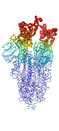
| - | [[Image:SpikeBFactor.png|250 px|left|rotate=180|thumb|Figure | + | [[Image:SpikeBFactor.png|250 px|left|rotate=180|thumb|Figure 2. Spike protein shown in "B-Factor"; depicting mobility and flexibility of different portions. Depicted in red are the most mobile, whilst dark blue are the least mobile. The 2 red portions depict RBDs, which correspond to 1-up and 2-up conformational states.]] |
<p>The <scene name='10/1075251/Spike_protein_closed_spacefill/4'>spike protein</scene> of SARS-COV-2 is a symmetric trimer featuring 3 spike glycoprotein chains (UNIPROT: [https://www.uniprot.org/uniprotkb/P0DTC2/entry P0DTC2]). Each monomer of the spike is called a spike glycoprotein, and the total assembly contains 2 main parts: The <scene name='10/1075251/Spike_protein_1-up_yang_s1/2'>S1</scene> and <scene name='10/1075251/Spike_protein_1-up_yang_s2/2'>S2</scene> subunits<ref name="Huang">DOI:10.1038/s41401-020-0485-4</ref>. However, the native spike protein does not exist in this state prior to infection. The protein is actually inactive initially, but is later activated by proteases cleaving the inactive S protein into its two active subunits<ref name="Huang">DOI:10.1038/s41401-020-0485-4</ref>. The S1 subunit contains the <scene name='10/1075251/Spike_protein_2-up_yang_s1_dom/2'>N-Terminal domain (NTD), C-Terminal Domain (CTD), and the Receptor Binding Domain (RBD)</scene>. The RBD is responsible for <scene name='10/1075251/Ace2_and_rbd/2'>binding to the ACE2 receptor</scene> on the surface of the target cell, as well as neutralizing antibodies. The NTD, CTD, and their relevant interfaces actually play much larger roles in the binding of the spike protein to ACE2 than the RBD does due to their larger surface areas<ref name="Huang">DOI:10.1038/s41401-020-0485-4</ref>. The S2 subunit is responsible for viral fusion and entry. Once bound to ACE2, and after the different domains in S2 have anchored to the membrane as well as delivered the viral envelope, the S2 subunit then changes conformation from the pre-hairpin to <scene name='10/1075251/Spike_protein_postfusion/2'>postfusion-hairpin</scene> conformation<ref name="Huang">DOI:10.1038/s41401-020-0485-4</ref>. The S2 subunit contains a fusion peptide domain (FP), heptapeptide repeat sequences 1 and 2 (HR1 & HR2), TM domain, and cytoplasmic fusion domain (CT). Full information about the location and structures of these domains within the S2 subunit can be found in references 1 and 3<ref name="Huang">DOI:10.1038/s41401-020-0485-4</ref><ref name="Zhang">PMID:34534731</ref>. For the purpose of this article about the minibinders, attention will be directed to the S1 subunit and its binding properties with ACE2. | <p>The <scene name='10/1075251/Spike_protein_closed_spacefill/4'>spike protein</scene> of SARS-COV-2 is a symmetric trimer featuring 3 spike glycoprotein chains (UNIPROT: [https://www.uniprot.org/uniprotkb/P0DTC2/entry P0DTC2]). Each monomer of the spike is called a spike glycoprotein, and the total assembly contains 2 main parts: The <scene name='10/1075251/Spike_protein_1-up_yang_s1/2'>S1</scene> and <scene name='10/1075251/Spike_protein_1-up_yang_s2/2'>S2</scene> subunits<ref name="Huang">DOI:10.1038/s41401-020-0485-4</ref>. However, the native spike protein does not exist in this state prior to infection. The protein is actually inactive initially, but is later activated by proteases cleaving the inactive S protein into its two active subunits<ref name="Huang">DOI:10.1038/s41401-020-0485-4</ref>. The S1 subunit contains the <scene name='10/1075251/Spike_protein_2-up_yang_s1_dom/2'>N-Terminal domain (NTD), C-Terminal Domain (CTD), and the Receptor Binding Domain (RBD)</scene>. The RBD is responsible for <scene name='10/1075251/Ace2_and_rbd/2'>binding to the ACE2 receptor</scene> on the surface of the target cell, as well as neutralizing antibodies. The NTD, CTD, and their relevant interfaces actually play much larger roles in the binding of the spike protein to ACE2 than the RBD does due to their larger surface areas<ref name="Huang">DOI:10.1038/s41401-020-0485-4</ref>. The S2 subunit is responsible for viral fusion and entry. Once bound to ACE2, and after the different domains in S2 have anchored to the membrane as well as delivered the viral envelope, the S2 subunit then changes conformation from the pre-hairpin to <scene name='10/1075251/Spike_protein_postfusion/2'>postfusion-hairpin</scene> conformation<ref name="Huang">DOI:10.1038/s41401-020-0485-4</ref>. The S2 subunit contains a fusion peptide domain (FP), heptapeptide repeat sequences 1 and 2 (HR1 & HR2), TM domain, and cytoplasmic fusion domain (CT). Full information about the location and structures of these domains within the S2 subunit can be found in references 1 and 3<ref name="Huang">DOI:10.1038/s41401-020-0485-4</ref><ref name="Zhang">PMID:34534731</ref>. For the purpose of this article about the minibinders, attention will be directed to the S1 subunit and its binding properties with ACE2. | ||
Revision as of 03:11, 15 April 2025
| This Sandbox is Reserved from March 18 through September 1, 2025 for use in the course CH462 Biochemistry II taught by R. Jeremy Johnson and Mark Macbeth at the Butler University, Indianapolis, USA. This reservation includes Sandbox Reserved 1828 through Sandbox Reserved 1846. |
To get started:
More help: Help:Editing |
SARS-COV2 Minibinders
| |||||||||||
Color Key
■ -> ACE2
■ -> Spike RBD
■ -> AHB2
■ -> LCB1
■ -> LCB3
References
[1] [2] [3] [4] [6] [5] [7] [8] [9]
- ↑ 1.00 1.01 1.02 1.03 1.04 1.05 1.06 1.07 1.08 1.09 1.10 1.11 1.12 1.13 1.14 1.15 1.16 1.17 Cao L, Goreshnik I, Coventry B, Case JB, Miller L, Kozodoy L, Chen RE, Carter L, Walls AC, Park YJ, Strauch EM, Stewart L, Diamond MS, Veesler D, Baker D. De novo design of picomolar SARS-CoV-2 miniprotein inhibitors. Science. 2020 Oct 23;370(6515):426-431. PMID:32907861 doi:10.1126/science.abd9909
- ↑ 2.0 2.1 2.2 2.3 2.4 Case JB, Chen RE, Cao L, Ying B, Winkler ES, Johnson M, Goreshnik I, Pham MN, Shrihari S, Kafai NM, Bailey AL, Xie X, Shi PY, Ravichandran R, Carter L, Stewart L, Baker D, Diamond MS. Ultrapotent miniproteins targeting the SARS-CoV-2 receptor-binding domain protect against infection and disease. Cell Host Microbe. 2021 Jul 14;29(7):1151-1161.e5. PMID:34192518 doi:10.1016/j.chom.2021.06.008
- ↑ 3.0 3.1 Sang P, Chen YQ, Liu MT, Wang YT, Yue T, Li Y, Yin YR, Yang LQ. Electrostatic Interactions Are the Primary Determinant of the Binding Affinity of SARS-CoV-2 Spike RBD to ACE2: A Computational Case Study of Omicron Variants. Int J Mol Sci. 2022 Nov 26;23(23):14796. PMID:36499120 doi:10.3390/ijms232314796
- ↑ 4.00 4.01 4.02 4.03 4.04 4.05 4.06 4.07 4.08 4.09 4.10 4.11 4.12 Huang Y, Yang C, Xu XF, Xu W, Liu SW. Structural and functional properties of SARS-CoV-2 spike protein: potential antivirus drug development for COVID-19. Acta Pharmacol Sin. 2020 Sep;41(9):1141-1149. doi: 10.1038/s41401-020-0485-4., Epub 2020 Aug 3. PMID:32747721 doi:http://dx.doi.org/10.1038/s41401-020-0485-4
- ↑ 5.0 5.1 5.2 5.3 Zhang J, Xiao T, Cai Y, Chen B. Structure of SARS-CoV-2 spike protein. Curr Opin Virol. 2021 Oct;50:173-182. PMID:34534731 doi:10.1016/j.coviro.2021.08.010
- ↑ 6.0 6.1 6.2 Yuan Y, Cao D, Zhang Y, Ma J, Qi J, Wang Q, Lu G, Wu Y, Yan J, Shi Y, Zhang X, Gao GF. Cryo-EM structures of MERS-CoV and SARS-CoV spike glycoproteins reveal the dynamic receptor binding domains. Nat Commun. 2017 Apr 10;8:15092. doi: 10.1038/ncomms15092. PMID:28393837 doi:http://dx.doi.org/10.1038/ncomms15092
- ↑ 7.0 7.1 7.2 Kuba K, Yamaguchi T, Penninger JM. Angiotensin-Converting Enzyme 2 (ACE2) in the Pathogenesis of ARDS in COVID-19. Front Immunol. 2021 Dec 22;12:732690. PMID:35003058 doi:10.3389/fimmu.2021.732690
- ↑ 8.0 8.1 Kuba K, Imai Y, Rao S, Gao H, Guo F, Guan B, Huan Y, Yang P, Zhang Y, Deng W, Bao L, Zhang B, Liu G, Wang Z, Chappell M, Liu Y, Zheng D, Leibbrandt A, Wada T, Slutsky AS, Liu D, Qin C, Jiang C, Penninger JM. A crucial role of angiotensin converting enzyme 2 (ACE2) in SARS coronavirus-induced lung injury. Nat Med. 2005 Aug;11(8):875-9. PMID:16007097 doi:10.1038/nm1267
- ↑ 9.0 9.1 Valetti F, Gilardi G. Improvement of biocatalysts for industrial and environmental purposes by saturation mutagenesis. Biomolecules. 2013 Oct 8;3(4):778-811. PMID:24970191 doi:10.3390/biom3040778


