This old version of Proteopedia is provided for student assignments while the new version is undergoing repairs. Content and edits done in this old version of Proteopedia after March 1, 2026 will eventually be lost when it is retired in about June of 2026.
Apply for new accounts at the new Proteopedia. Your logins will work in both the old and new versions.
2dfx
From Proteopedia
| Line 1: | Line 1: | ||
| - | [[Image:2dfx. | + | {{Seed}} |
| + | [[Image:2dfx.png|left|200px]] | ||
<!-- | <!-- | ||
| Line 9: | Line 10: | ||
{{STRUCTURE_2dfx| PDB=2dfx | SCENE= }} | {{STRUCTURE_2dfx| PDB=2dfx | SCENE= }} | ||
| - | + | ===Crystal structure of the carboxy terminal domain of colicin E5 complexed with its inhibitor=== | |
| - | + | <!-- | |
| - | + | The line below this paragraph, {{ABSTRACT_PUBMED_17099236}}, adds the Publication Abstract to the page | |
| + | (as it appears on PubMed at http://www.pubmed.gov), where 17099236 is the PubMed ID number. | ||
| + | --> | ||
| + | {{ABSTRACT_PUBMED_17099236}} | ||
==About this Structure== | ==About this Structure== | ||
| Line 30: | Line 34: | ||
[[Category: Alpha/beta protein]] | [[Category: Alpha/beta protein]] | ||
[[Category: Protein-inhibitor protein complex]] | [[Category: Protein-inhibitor protein complex]] | ||
| - | ''Page seeded by [http://oca.weizmann.ac.il/oca OCA ] on | + | |
| + | ''Page seeded by [http://oca.weizmann.ac.il/oca OCA ] on Mon Jul 28 09:29:52 2008'' | ||
Revision as of 06:29, 28 July 2008
| |||||||||
| 2dfx, resolution 1.90Å () | |||||||||
|---|---|---|---|---|---|---|---|---|---|
| Related: | 2djh | ||||||||
| |||||||||
| |||||||||
| |||||||||
| Resources: | FirstGlance, OCA, PDBsum, RCSB | ||||||||
| Coordinates: | save as pdb, mmCIF, xml | ||||||||
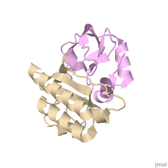

Crystal structure of the carboxy terminal domain of colicin E5 complexed with its inhibitor
Template:ABSTRACT PUBMED 17099236
About this Structure
2DFX is a Protein complex structure of sequences from Escherichia coli. Full crystallographic information is available from OCA.
Reference
Structural basis for sequence-dependent recognition of colicin E5 tRNase by mimicking the mRNA-tRNA interaction., Yajima S, Inoue S, Ogawa T, Nonaka T, Ohsawa K, Masaki H, Nucleic Acids Res. 2006;34(21):6074-82. Epub 2006 Nov 11. PMID:17099236
Page seeded by OCA on Mon Jul 28 09:29:52 2008
