This old version of Proteopedia is provided for student assignments while the new version is undergoing repairs. Content and edits done in this old version of Proteopedia after March 1, 2026 will eventually be lost when it is retired in about June of 2026.
Apply for new accounts at the new Proteopedia. Your logins will work in both the old and new versions.
1czf
From Proteopedia
(New page: 200px<br /><applet load="1czf" size="450" color="white" frame="true" align="right" spinBox="true" caption="1czf, resolution 1.68Å" /> '''ENDO-POLYGALACTURONA...) |
|||
| Line 1: | Line 1: | ||
| - | [[Image:1czf.gif|left|200px]]<br /><applet load="1czf" size=" | + | [[Image:1czf.gif|left|200px]]<br /><applet load="1czf" size="350" color="white" frame="true" align="right" spinBox="true" |
caption="1czf, resolution 1.68Å" /> | caption="1czf, resolution 1.68Å" /> | ||
'''ENDO-POLYGALACTURONASE II FROM ASPERGILLUS NIGER'''<br /> | '''ENDO-POLYGALACTURONASE II FROM ASPERGILLUS NIGER'''<br /> | ||
==Overview== | ==Overview== | ||
| - | Polygalacturonases specifically hydrolyze polygalacturonate, a major | + | Polygalacturonases specifically hydrolyze polygalacturonate, a major constituent of plant cell wall pectin. To understand the catalytic mechanism and substrate and product specificity of these enzymes, we have solved the x-ray structure of endopolygalacturonase II of Aspergillus niger and we have carried out site-directed mutagenesis studies. The enzyme folds into a right-handed parallel beta-helix with 10 complete turns. The beta-helix is composed of four parallel beta-sheets, and has one very small alpha-helix near the N terminus, which shields the enzyme's hydrophobic core. Loop regions form a cleft on the exterior of the beta-helix. Site-directed mutagenesis of Asp(180), Asp(201), Asp(202), His(223), Arg(256), and Lys(258), which are located in this cleft, results in a severe reduction of activity, demonstrating that these residues are important for substrate binding and/or catalysis. The juxtaposition of the catalytic residues differs from that normally encountered in inverting glycosyl hydrolases. A comparison of the endopolygalacturonase II active site with that of the P22 tailspike rhamnosidase suggests that Asp(180) and Asp(202) activate the attacking nucleophilic water molecule, while Asp(201) protonates the glycosidic oxygen of the scissile bond. |
==About this Structure== | ==About this Structure== | ||
| - | 1CZF is a [http://en.wikipedia.org/wiki/Single_protein Single protein] structure of sequence from [http://en.wikipedia.org/wiki/Aspergillus_niger Aspergillus niger] with NAG and ZN as [http://en.wikipedia.org/wiki/ligands ligands]. Active as [http://en.wikipedia.org/wiki/Polygalacturonase Polygalacturonase], with EC number [http://www.brenda-enzymes.info/php/result_flat.php4?ecno=3.2.1.15 3.2.1.15] Full crystallographic information is available from [http:// | + | 1CZF is a [http://en.wikipedia.org/wiki/Single_protein Single protein] structure of sequence from [http://en.wikipedia.org/wiki/Aspergillus_niger Aspergillus niger] with <scene name='pdbligand=NAG:'>NAG</scene> and <scene name='pdbligand=ZN:'>ZN</scene> as [http://en.wikipedia.org/wiki/ligands ligands]. Active as [http://en.wikipedia.org/wiki/Polygalacturonase Polygalacturonase], with EC number [http://www.brenda-enzymes.info/php/result_flat.php4?ecno=3.2.1.15 3.2.1.15] Full crystallographic information is available from [http://oca.weizmann.ac.il/oca-bin/ocashort?id=1CZF OCA]. |
==Reference== | ==Reference== | ||
| Line 14: | Line 14: | ||
[[Category: Polygalacturonase]] | [[Category: Polygalacturonase]] | ||
[[Category: Single protein]] | [[Category: Single protein]] | ||
| - | [[Category: Dijkstra, B | + | [[Category: Dijkstra, B W.]] |
| - | [[Category: Kalk, K | + | [[Category: Kalk, K H.]] |
| - | [[Category: Santen, Y | + | [[Category: Santen, Y van.]] |
[[Category: NAG]] | [[Category: NAG]] | ||
[[Category: ZN]] | [[Category: ZN]] | ||
[[Category: beta helix]] | [[Category: beta helix]] | ||
| - | ''Page seeded by [http:// | + | ''Page seeded by [http://oca.weizmann.ac.il/oca OCA ] on Thu Feb 21 12:11:16 2008'' |
Revision as of 10:11, 21 February 2008
|
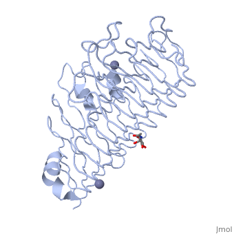
ENDO-POLYGALACTURONASE II FROM ASPERGILLUS NIGER
Overview
Polygalacturonases specifically hydrolyze polygalacturonate, a major constituent of plant cell wall pectin. To understand the catalytic mechanism and substrate and product specificity of these enzymes, we have solved the x-ray structure of endopolygalacturonase II of Aspergillus niger and we have carried out site-directed mutagenesis studies. The enzyme folds into a right-handed parallel beta-helix with 10 complete turns. The beta-helix is composed of four parallel beta-sheets, and has one very small alpha-helix near the N terminus, which shields the enzyme's hydrophobic core. Loop regions form a cleft on the exterior of the beta-helix. Site-directed mutagenesis of Asp(180), Asp(201), Asp(202), His(223), Arg(256), and Lys(258), which are located in this cleft, results in a severe reduction of activity, demonstrating that these residues are important for substrate binding and/or catalysis. The juxtaposition of the catalytic residues differs from that normally encountered in inverting glycosyl hydrolases. A comparison of the endopolygalacturonase II active site with that of the P22 tailspike rhamnosidase suggests that Asp(180) and Asp(202) activate the attacking nucleophilic water molecule, while Asp(201) protonates the glycosidic oxygen of the scissile bond.
About this Structure
1CZF is a Single protein structure of sequence from Aspergillus niger with and as ligands. Active as Polygalacturonase, with EC number 3.2.1.15 Full crystallographic information is available from OCA.
Reference
1.68-A crystal structure of endopolygalacturonase II from Aspergillus niger and identification of active site residues by site-directed mutagenesis., van Santen Y, Benen JA, Schroter KH, Kalk KH, Armand S, Visser J, Dijkstra BW, J Biol Chem. 1999 Oct 22;274(43):30474-80. PMID:10521427
Page seeded by OCA on Thu Feb 21 12:11:16 2008
