This old version of Proteopedia is provided for student assignments while the new version is undergoing repairs. Content and edits done in this old version of Proteopedia after March 1, 2026 will eventually be lost when it is retired in about June of 2026.
Apply for new accounts at the new Proteopedia. Your logins will work in both the old and new versions.
Apply for new accounts at the new Proteopedia. Your logins will work in both the old and new versions.
Sandbox2qc8
From Proteopedia
(Difference between revisions)
| Line 1: | Line 1: | ||
{{STRUCTURE_2qc8 | PDB=2qc8 | SCENE= }} | {{STRUCTURE_2qc8 | PDB=2qc8 | SCENE= }} | ||
| + | |||
| + | =Exercises= | ||
Click here to view <scene name='Practice_Page/Exercise1/1'>Exercise 1 Results</scene>. | Click here to view <scene name='Practice_Page/Exercise1/1'>Exercise 1 Results</scene>. | ||
| Line 9: | Line 11: | ||
Click here to view <scene name='Sandbox2qc8/Exercise_4/1'>Exercise 4 Results</scene>. | Click here to view <scene name='Sandbox2qc8/Exercise_4/1'>Exercise 4 Results</scene>. | ||
| + | |||
| + | =Outline= | ||
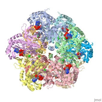
Glutamine synthetase is composed of 10 <scene name='Sandbox2qc8/Secondary_structure/1'>identical subunits</scene>. | Glutamine synthetase is composed of 10 <scene name='Sandbox2qc8/Secondary_structure/1'>identical subunits</scene>. | ||
| Line 27: | Line 31: | ||
=References= | =References= | ||
<references/> | <references/> | ||
| + | |||
| + | == Headline text == | ||
Revision as of 03:06, 8 December 2008
| |||||||||
| 2qc8, resolution 2.60Å () | |||||||||
|---|---|---|---|---|---|---|---|---|---|
| Ligands: | , , , | ||||||||
| Gene: | GLUL, GLNS (Homo sapiens) | ||||||||
| Activity: | Glutamate--ammonia ligase, with EC number 6.3.1.2 | ||||||||
| Related: | 2ojw | ||||||||
| |||||||||
| |||||||||
| Resources: | FirstGlance, OCA, RCSB, PDBsum | ||||||||
| Coordinates: | save as pdb, mmCIF, xml | ||||||||

Contents |
Exercises
Click here to view .
Click here to view .
Click here to view .
Click here to view .
Outline
Glutamine synthetase is composed of 10 .
Each subunit has an exposed NH2 terminus and buried COOH terminus as part of a helical thong. [1]
Each subunit is composed predominantly of 12 and 12 , as well as a .
The beta sheets are arranged into two separate partial beta barrels, one of which encompasses the ligand complex.
The active site within the secondary structure can be called a "bifunnel," providing access to ATP and glutamate at opposing ends.[2]
The ligands present are Cl, Mn, Adenosine Diphosphate, and L-Methionine-S-Sulfoximine Phosphate.
References
- ↑ Yamashita, M., et al.,Refined Atomic Model of Glutamine Synthetase at 3.5A Resolution, The Journal of Biological Chemistry, 1989, 17681-17690.
- ↑ Eisenberg, D., et al., Structure-function relationships of glutamine synthetases, Biochimica et Biophysica Acta 1477 (2000), 122-145.
