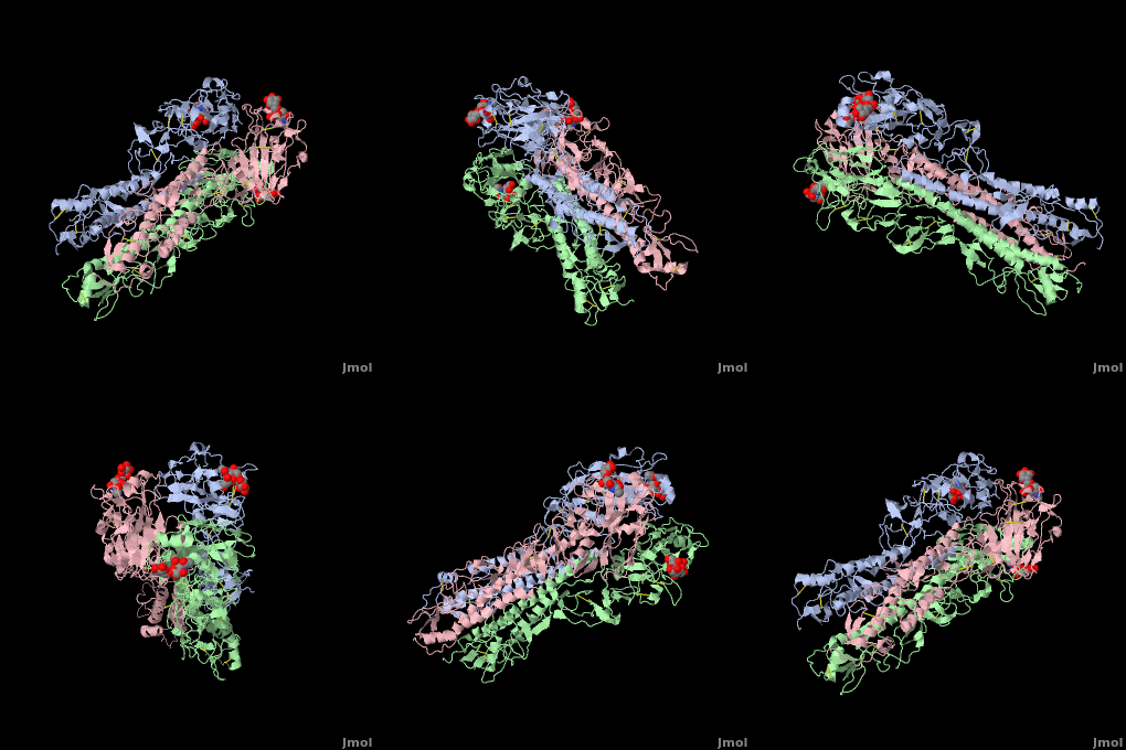
Hemagglutinin
From Proteopedia
(→2WR2) |
|||
| Line 1: | Line 1: | ||
==XWR2== | ==XWR2== | ||
| - | '''Generalities''' | + | '''[[Generalities]]''' |
Hemagglutinins (HA) are one of the two antigenic Glycoproteins inserted into the influenza virus membrane. There are at least 16 different forms of HA Antigens classified from H1 to H16. H1, H2, H3 are specific of Humans Influenza . | Hemagglutinins (HA) are one of the two antigenic Glycoproteins inserted into the influenza virus membrane. There are at least 16 different forms of HA Antigens classified from H1 to H16. H1, H2, H3 are specific of Humans Influenza . | ||
| Line 22: | Line 22: | ||
His 183, Tyr 98, Glu 190, Ser 136 form Hydrogen bound with the receptor and the Leu 194 and the Trp 153 make have a potential to make Van der Waals contact with the hydrophobic portion of the receptor. | His 183, Tyr 98, Glu 190, Ser 136 form Hydrogen bound with the receptor and the Leu 194 and the Trp 153 make have a potential to make Van der Waals contact with the hydrophobic portion of the receptor. | ||
| - | '''2WR2''' | + | '''[[2WR2]]''' |
To begin with, this code corresponds to the structure of influenza H2 Avian Hemagglutinin with avian receptor. | To begin with, this code corresponds to the structure of influenza H2 Avian Hemagglutinin with avian receptor. | ||
The difference between the Avian H2 and Human H2 is due to a mutation on two amino acids: | The difference between the Avian H2 and Human H2 is due to a mutation on two amino acids: | ||
| Line 33: | Line 33: | ||
| - | {{ | + | {{STRUCTURE_ | PDB=2WR2 | SCENE=2WR2 }} |
Revision as of 08:34, 31 October 2009
XWR2
Hemagglutinins (HA) are one of the two antigenic Glycoproteins inserted into the influenza virus membrane. There are at least 16 different forms of HA Antigens classified from H1 to H16. H1, H2, H3 are specific of Humans Influenza . HA have two main functions which are implied in the viruses’ infection cycle: 1) On the target cell, HA bind the receptor which is composed by Sialic acid, allowing the virus/cell interaction. 2) HA induce the fusion between the host cell and the virus which can entry into the cytoplasm. Because the HA are the major Antigens of the Virus, Antibodies recognized them and for this reason HA often change.
HA is a homotrimeric integral membrane glycoprotein. Each monomer is synthesized like a single polypeptide chain almost 550 amino acids. This precursor is then glycosilated and cleaved into two smaller polypeptides by removal of Arginin 329; at the same time, a conformational change occurs in the monomer.
The HA1 and HA2 subunits are covalently attached by a disulfide bound from HA1 position 14 to HA2 position 137. These two chains form one monomer, and the noncovalentely association of three monomers forms one hemagglutinin molecule: (HA1+HA2)3. It is principally stabilized by packing of the alpha-helixes. All molecules are 135 angstrom long.
The HA1 subunit (328 amino acids) has a globular form outside the virus’ membrane: it is composed by an eight-stranded beta-sheet, associated with a little alpha-helix, separating the strands 3 and 4. The amino acids of this alpha-helix, and some others around it, included in the beta-sheet, compose the binding site for the receptor’s sialic acid. Thus, for one molecule of hemagglutinin, there are three binding sites to the host cell’s receptor.
The HA2 subunit (221 amino acids) has a hair spin structure composed by two antiparallel alpha-helixes. One of these belongs to the longest alpha-helixes in globular protein: it is 50 angstrom long. The hydrophobic N-terminus part of HA2, called fusion peptide, is close to a protease’s cleavage site: this protease is implied in the virus’ entry in the host cell.
HA1 and HA2 are bound each other by a disulfide bind. The three long alpha-helixes (of the three HA2) are coiled-coil to form a central region of 40 angstrom, and thank to the hydrophobic amino acids and those which form salt bridges bound, we obtain a HA stabilized.
Five amino acids compose the catalytic site (Tyr 98, His 183, Glu 190, Trp 153, Leu 194) which are identical in the all different HA. The catalytic structure is stabilized by other conserved amino acid which cannot interact with the receptor (Cys 97, Pro 99, Cis139, Pro 147, Tyr 195, Arg 229). His 183, Tyr 98, Glu 190, Ser 136 form Hydrogen bound with the receptor and the Leu 194 and the Trp 153 make have a potential to make Van der Waals contact with the hydrophobic portion of the receptor.
2WR2 To begin with, this code corresponds to the structure of influenza H2 Avian Hemagglutinin with avian receptor. The difference between the Avian H2 and Human H2 is due to a mutation on two amino acids: - At the position 226, Avian Hemagglutinin have a Glutamine residue whereas the Human one has Leucine - At the position 228, there is a Glycin for Avian HA whereas a Serine for Human HA. With these two mutations, the human H2 cannot bind the avian receptor. But avian virus can bind to the human receptor.
The avian H2 can bind to the human receptor because of Hydrogen bonds networks created by Gln 226 and by Asn 186. The complex between H2 HA
| |||||||
| {{{CAPTION}}} | |||||||
|---|---|---|---|---|---|---|---|
| Coordinates: | save as pdb, mmCIF, xml | ||||||
Proteopedia Page Contributors and Editors (what is this?)
Julien Madouasse, Michal Harel, David Canner, Mark Hoelzer, Ann Taylor, Alexander Berchansky, Joel L. Sussman

