User:Claire Roudot/Sandbox 1
From Proteopedia
| Line 3: | Line 3: | ||
Prions are proteins Prp-sc which have a different domain conformation than the natural protein Prp-c. | Prions are proteins Prp-sc which have a different domain conformation than the natural protein Prp-c. | ||
The prion form of HET-s plays a role in heterokaryon incompatibility, a fungal self/nonself recognition phenomenon that prevents different form of parasitism. | The prion form of HET-s plays a role in heterokaryon incompatibility, a fungal self/nonself recognition phenomenon that prevents different form of parasitism. | ||
| + | [[Image:200px-2rnm.png]] | ||
| + | |||
Structure: | Structure: | ||
Revision as of 08:26, 12 November 2009
|
Prions are often implicated in disease such as Creutzeld Jakob disease in humans, it can be also found in yeast and filamentous fungi.
Prions are proteins Prp-sc which have a different domain conformation than the natural protein Prp-c.
The prion form of HET-s plays a role in heterokaryon incompatibility, a fungal self/nonself recognition phenomenon that prevents different form of parasitism.

Structure:
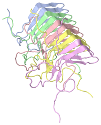
The global organization of the HET-s (218-289) fibril is a left handed β solenoid with two windings per molecule. The studies of this part of molecule have been done with NMR.
HET-s (218-289) is formed by four β strands, each strand is formed two parallel β sheet with short HN-Hα contacts (3,0A). Three strands constitute the core of the fibril. The last strand is outside the core and is formed by β2b and β4b. The segments β1a and β1b (β3a and β3b) have an approximately rectangular kink between them thanks a connection by a two-residue β arc, changing the inside-outside pattern of side chains. The connection between β1b and β2a (and similarly, between β3b and β4a) is linked by a three-residue β arc, allowing for the orientation change of the polypeptide backbone by ~ 150°. The β-sheet pattern is disrupted between β2a and β2b, leading to ~90° arcs. β1-β2 and β3-β4 are pseudo repeats and linked by parallel intramolecular and intermolecular H bonds, as follows: β1a-β3a, β1b-β3b, β2a-β4a, β2b-β4b.
The first three β strands of each pseudo-repeat realized a triangular hydrophobic core, which is tightly packed, where almost exclusively hydrophobic residues are contained (Ala, Leu, Ile and Val). These strands created a very dense packing where water is quite exclude. In contrast, all charged residues face outside and are mostly located in arc β-arc regions, where the solvent accessibility is high. Three of them are arranged on top of each other such that charges are compensated (montrer sur figure) and the formation of salt-bridges becomes possible. Because the stacking is parallel, it may have intra- and intermolecular charge compensation This may explain why fibrils have high stability against denaturation by non-ionic urea at neutral pH, but are destabilized by urea at acidic or basic pH.
The structure of HET-s(218-289) with its overall β-helical fold is more complexed than that of peptide fibril- this complexity is often is soluble proteins. This complex alternance of pseudo-repeat (β1/β3 and β2/β4) permits the alternation of positive and negative charges along the fibril axis, and also having a molecule forming two turns of the solenoid.
The triangular hydrophobic core formed by three stands has some resemblance with β-solenoid structures of soluble proteins like filamentous hemagglutinin protein. In this case, the periodicity does not exist but he triangular core is quite similar. A β-solenoid fold has also been proposed for the prion state of human prion protein Prp (…..).
This well-organized structure of the HET-s prion fibrils is originally from the high order is these fibrils as well as the absence of polymorphism caused by different underlying molecular at physiological pH conditions. Indeed interaction between charges in the fibril gives a high stability, so polymorphism is excluding. The fibril structure of HET-s (218-289) along an axe amplified the well-defined structure of a functional amyloid.
