This old version of Proteopedia is provided for student assignments while the new version is undergoing repairs. Content and edits done in this old version of Proteopedia after March 1, 2026 will eventually be lost when it is retired in about June of 2026.
Apply for new accounts at the new Proteopedia. Your logins will work in both the old and new versions.
The Structure and Mechanism of Hexokinase
From Proteopedia
| Line 15: | Line 15: | ||
In the first reaction of glycolysis, the gamma-phosphoryl group of an ATP molecule is transferred to the oxygen at the C-6 of glucose. The magnesium ion is required as the reactive form of ATP is the complex with magnesium (II) ion. This step is a direct nucleophilic attack of the hydroxyl group on the terminal phosphoryl group of the ATP molecule. This produces glucose-6-phosphate and ADP. Hexokinase is the enzyme that catalyzes this phosphoryl group transfer. Hexokinase undergoes and induced-fit conformational change when it binds to glucose, which ultimately prevents the hydrolysis of ATP. It is also allosterically inhibited by physiological concentrations of its immediate product, glucose-6-phosphate. This is a mechanism by which the influx of substrate into the glycolytic pathway is controlled. | In the first reaction of glycolysis, the gamma-phosphoryl group of an ATP molecule is transferred to the oxygen at the C-6 of glucose. The magnesium ion is required as the reactive form of ATP is the complex with magnesium (II) ion. This step is a direct nucleophilic attack of the hydroxyl group on the terminal phosphoryl group of the ATP molecule. This produces glucose-6-phosphate and ADP. Hexokinase is the enzyme that catalyzes this phosphoryl group transfer. Hexokinase undergoes and induced-fit conformational change when it binds to glucose, which ultimately prevents the hydrolysis of ATP. It is also allosterically inhibited by physiological concentrations of its immediate product, glucose-6-phosphate. This is a mechanism by which the influx of substrate into the glycolytic pathway is controlled. | ||
| - | Five loops exist that exhibit conformational change that cannot be represented by rigid body movements. The specific residues involved in the conformational change include Lys 621 which binds to glucose and a second loop (residues 618-624) that interact with glucose. Both of these loops relax together to new conformations in the absence of glucose. Loop 532-537 which bind G6P, also relaxes in the absence of the inhibitor. Thr 536 hydrogen bonds with the 6-phosphoryl group of the inhibitor in the glucose/G6P dimer, and may be a factor in the tight binding of G6P by hexokinase. Three rigid-body transformations, which convert the glucose/Pi conformer into the glucose/G6P conformer, prompt the belief in existence of hinges in the polypeptide chain of hexokinase. One hinge spans from residues 462 to 469 of the transition helix between the N and C terminal halves. These residues are hydrophobic, being exposed to solvent and free of side-chain hydrogen bonds to other elements of the protein. Three additional hinges allow the relative movement of the small and large domains of the C-terminal half. The existance of a salt link between Arg 539 and Asp 895 of the open conformation of the C-terminal half breaks in the closed conformation. Arg 539 is essential for catalysis interacting with a phosphoryl group of ATP. | + | Five loops exist that exhibit conformational change that cannot be represented by rigid body movements. The specific residues involved in the conformational change include Lys 621 which binds to glucose and a second loop (residues 618-624) that interact with glucose. Both of these loops relax together to new conformations in the absence of glucose. Loop 532-537 which bind G6P, also relaxes in the absence of the inhibitor. Thr 536 hydrogen bonds with the 6-phosphoryl group of the inhibitor in the glucose/G6P dimer, and may be a factor in the tight binding of G6P by hexokinase. Three rigid-body transformations, which convert the glucose/Pi conformer into the glucose/G6P conformer, prompt the belief in existence of hinges in the polypeptide chain of hexokinase. One hinge spans from residues 462 to 469 of the transition helix between the N and C terminal halves. These residues are hydrophobic, being exposed to solvent and free of side-chain hydrogen bonds to other elements of the protein. Three additional hinges allow the relative movement of the small and large domains of the C-terminal half. The existance of a salt link between Arg 539 and Asp 895 of the open conformation of the C-terminal half breaks in the closed conformation. Arg 539 is essential for catalysis interacting with a phosphoryl group of ATP <ref>PMID:7048063</ref>. |
Revision as of 19:47, 1 March 2010
|
The Structure and Mechanism of Hexokinase
A hexokinase is an enzyme that phosphorylates a six-carbon sugar, a hexose, to a hexose phosphate. In most tissues and organisms, glucose is the most important substrate of hexokinases, and glucose 6-phosphate the most important product. Hexokinases have been found in every organism checked, ranging from bacteria, yeast, and plants, to humans and other vertebrates. They are categorized as actin fold proteins, sharing a common ATP binding site core surrounded by more variable sequences that determine substrate affinities and other properties. Several hexokinase isoforms or isozymes providing different functions can occur in a single species.
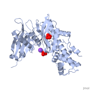
Hexokinase Structure: Hexokinase contains 17 alpha helices and 11 beta sheets. The tertiary structure of hexokinase includes an open alpha/beta sheet. There is a large amount of variation associated with this structure. It is composed of five beta sheets and three alpha helices. In this open alph/beta sheet four of the beta sheets are parallel and one is in the anitparallel directions. The alpha helices and beta loops connect the beta sheets to produce this open alpha/beta sheet. The crevice indicates the ATP-binding domain of this glycolytic enzyme.
The hexokinase molecule has two distinct conformations, and , and the conformational fluctuation between the two states involves relative motion of the two domains or two halves of the protein. The existance of a salt link between Arg 539 and Asp 895 of the open conformation of the C-terminal half breaks in the closed conformation. Arg 539 is essential for catalysis interacting with a phosphory group of ATP. In the open conformation, the molecule has a low affinity for both the glucose molecule and the ATP molecule. The binding of one of the molecules, say glucose, shifts the equilibrium to the closed conformation of the protein, which has a higher affinity for ATP because now the ATP binding site has the correct conformation to accommodate ATP. By the same reasoning, if ATP were to bind first, that would also shift the equilibrium to the closed conformation and hence increase the affinity for glucose. Therefore the binding of glucose and ATP are coupled and this kind of conformational coupling makes hexokinase an allosteric protein. They are categorized as actin fold proteins, sharing a common ATP binding site core surrounded by more variable sequences that determine substrate affinities and other properties.
Mechanism of Hexokinase: In the first reaction of glycolysis, the gamma-phosphoryl group of an ATP molecule is transferred to the oxygen at the C-6 of glucose. The magnesium ion is required as the reactive form of ATP is the complex with magnesium (II) ion. This step is a direct nucleophilic attack of the hydroxyl group on the terminal phosphoryl group of the ATP molecule. This produces glucose-6-phosphate and ADP. Hexokinase is the enzyme that catalyzes this phosphoryl group transfer. Hexokinase undergoes and induced-fit conformational change when it binds to glucose, which ultimately prevents the hydrolysis of ATP. It is also allosterically inhibited by physiological concentrations of its immediate product, glucose-6-phosphate. This is a mechanism by which the influx of substrate into the glycolytic pathway is controlled.
Five loops exist that exhibit conformational change that cannot be represented by rigid body movements. The specific residues involved in the conformational change include Lys 621 which binds to glucose and a second loop (residues 618-624) that interact with glucose. Both of these loops relax together to new conformations in the absence of glucose. Loop 532-537 which bind G6P, also relaxes in the absence of the inhibitor. Thr 536 hydrogen bonds with the 6-phosphoryl group of the inhibitor in the glucose/G6P dimer, and may be a factor in the tight binding of G6P by hexokinase. Three rigid-body transformations, which convert the glucose/Pi conformer into the glucose/G6P conformer, prompt the belief in existence of hinges in the polypeptide chain of hexokinase. One hinge spans from residues 462 to 469 of the transition helix between the N and C terminal halves. These residues are hydrophobic, being exposed to solvent and free of side-chain hydrogen bonds to other elements of the protein. Three additional hinges allow the relative movement of the small and large domains of the C-terminal half. The existance of a salt link between Arg 539 and Asp 895 of the open conformation of the C-terminal half breaks in the closed conformation. Arg 539 is essential for catalysis interacting with a phosphoryl group of ATP [1].
Glucokinase, an Isoenzyme of Hexokinase
Glucokinase (hexokinase D) is a monomeric cytoplasmic enzyme found in the liver and pancreas but can also be found in the gut and brain. It serves to regulate glucose levels in these organs. Glucokinase uses phosphorylation to increase the metabolism of glucose. Glucokinase is a hexokinase isoenzyme. All hexokinases are capable of prompting the first step of glycogen synthesis and glycolysis, the phosphorylation of glucose to glucose-6-phosphate (G6P).
Glucokinase vs. Other Hexokinases: Glucokinase is unique from other hexokinase in kinetic properties and is coded by a different gene. The difference of glucokinase from the other hexokinases is that glucokinase has a lower affinity, thus a higher Km, for glucose. The reduced affinity for glucose allows the activity of glucokinase to differ under physiological conditions according to the amount of glucose present. Essentially, this means that it operates only when serum glucose levels are high. High glucose is the signal to store glucose. Other tissues need to use glucose at lower serum levels and thus use the higher affinity (lower Km) hexokinase. Also, G6P inhibits hexokinase. This is simple "product inhibition". If the cell is not using up the G6P that it is making, then it should stop making it. G6P does not inhibit glucokinase. This allows it to remain active in storing as much glucose as possible in the presence of high glucose levels.
Role in Organ Systems: In the liver glucokinase increases the synthesis of glycogen and is the first step in glycolysis, the main producer of ATP in the body. Glucokinase is responsible for phospohorylating the majority of glucose in the liver and pancreas. Glucokinase only binds to and phosphorylates glucose when levels are higher than normal blood glucose level, allowing it to maintain constant glucose levels[2]. By phosphorylating glucose, glucokinase creates glucose 6-phosphate. Glucose 6-phosphate can then be used by the liver through the glycolytic pathway. Along with this process in the liver, glucokinase also facilitates glycogen synthesis. Through this the majority of the body's glucose is stored. Glucose 6-phosphate is also one of the starting materials of the TCA cycle which is responsible for the majority of ATP production in the body.
In the pancreas, a rise in glucose levels increases the activity of glucokinase causing an increase in glucose 6-phosphate. This causes the triggering of the beta cells to secret insulin[3]. Glucokinase is the first step in this reaction. Insulin then allows other cells in the body to take up glucose, actively lowering the glucose level
- ↑ Pollard-Knight D, Cornish-Bowden A. Mechanism of liver glucokinase. Mol Cell Biochem. 1982 Apr 30;44(2):71-80. PMID:7048063
- ↑ Kamata K, Mitsuya M, Nishimura T, Eiki J, Nagata Y. Structural basis for allosteric regulation of the monomeric allosteric enzyme human glucokinase. Structure. 2004 Mar;12(3):429-38. PMID:15016359 doi:10.1016/j.str.2004.02.005
- ↑ Postic C, Shiota M, Magnuson MA. Cell-specific roles of glucokinase in glucose homeostasis. Recent Prog Horm Res. 2001;56:195-217. PMID:11237213
Proteopedia Page Contributors and Editors (what is this?)
Kyle Schroering, Ann Taylor, Michal Harel, Drew McCaffrey, Alexander Berchansky, Joel L. Sussman, Cody Leatherman, David Canner
