Sandbox 160
From Proteopedia
| Line 11: | Line 11: | ||
Glyceraldehyde 3-Phosphate Dehydrogenase (GAPDH)has carefully been studied in a number of bacterial, parasitic and mammalian species and it has been found that it exists as homotetrameric protein <ref name="reference 1"/>. Each subunit within the protein is 38,151Da (tetramer is 152.4 kDa)and contains seven alpha helices and two beta sheets one of which has seven strands and the other with eight<ref name="reference 1"/> <ref name="ref 5">PMID:15953771</ref>. Two anion binding sites have been found where the two phosphates involved in the reaction will be bound during catalysis. One site is labeled "Pi" and is the location where the inorganic phosphate involved will bind and the other has been labeled "Ps" which is where the C-3 phosphate of Gylceraldeyhde 3-Phosphate will bind <ref name="reference 1"/>. Further experimentation has shown that the "Ps" site has been conserved in numerous GAPDH complexes and that the former may involve two possible sites in which the second or new "Pi" site is located 2.9 Angstroms from the primary "Pi" site<ref name="reference 1"/>. | Glyceraldehyde 3-Phosphate Dehydrogenase (GAPDH)has carefully been studied in a number of bacterial, parasitic and mammalian species and it has been found that it exists as homotetrameric protein <ref name="reference 1"/>. Each subunit within the protein is 38,151Da (tetramer is 152.4 kDa)and contains seven alpha helices and two beta sheets one of which has seven strands and the other with eight<ref name="reference 1"/> <ref name="ref 5">PMID:15953771</ref>. Two anion binding sites have been found where the two phosphates involved in the reaction will be bound during catalysis. One site is labeled "Pi" and is the location where the inorganic phosphate involved will bind and the other has been labeled "Ps" which is where the C-3 phosphate of Gylceraldeyhde 3-Phosphate will bind <ref name="reference 1"/>. Further experimentation has shown that the "Ps" site has been conserved in numerous GAPDH complexes and that the former may involve two possible sites in which the second or new "Pi" site is located 2.9 Angstroms from the primary "Pi" site<ref name="reference 1"/>. | ||
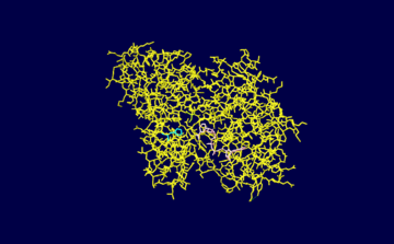
| - | The enzyme contains a functional NAD+ group which functions as a hydrogen acceptor during the course of the reaction which is bound to a Rossman fold. During the catalysis of glyceraldehyde 3-phosphate to 1,3-biphosphoglycerate a hydride ion is enzymatically transferred from the aldehyde group of glyceraldehyde 3-phosphate to the nicotinamide ring of NAD+ reducing it to NADH | + | The enzyme contains a functional NAD+ group which functions as a hydrogen acceptor during the course of the reaction which is bound to a Rossman fold. During the catalysis of glyceraldehyde 3-phosphate to 1,3-biphosphoglycerate a hydride ion is enzymatically transferred from the aldehyde group of glyceraldehyde 3-phosphate to the nicotinamide ring of NAD+ reducing it to NADH<ref name="reference 1">PMID:19243605 </ref>. The active site of GAPDH contains a cysteine (Cys149) residue which reacts with the glyceraldehyde 3-phosphate molecule through its -SH group. The substrate is covalently bound during the reaction through its aldehyde group to the -SH group of the cysteine residue and the resulting reaction produces a thiohemiacetal intermediate <ref name="reference 1"/>. Note that this reaction occurs through acid base catalysis with aid of a histidine residue (His176).The <scene name='Sandbox_160/Newscene/1'>active site</scene> of the molecule is illustrated to the right. |
A simplified illustration of the net reaction is as follows: | A simplified illustration of the net reaction is as follows: | ||
| Line 24: | Line 24: | ||
== Active Site in Detail == | == Active Site in Detail == | ||
| - | [[Image:1vc2 image.png | left|thumb|upright=2 |Figure 1. Illustration created using PDB software highlighting the NAD+ ligand and Cysteine and Histidine residues within the active site of 1vc2.]] Once Glyceraldehyde 3-phophate comes into contact with the active site it forms a hydrogen bond through its C2 hydroxyl group to Cys149N. The C1 hydroxyl group of the substrate binds to His176NE2<ref name="ref 4">PMID:10191140 </ref>. Additional hydrogen bonds to the phosphate group of the substrate from additional residues such as Thr1790G1, Arg231NH1(these two residues are not highlighted in the illustration below) along with N7N and 02'N of the NAD+ moeity (pink) help stabilize the molecule during the course of the reaction in the active site <ref name="ref 4"/>. The nicotinamide ring of the NAD+ ligand is responsible for orienting the hydrogen atom at C1 towards itself which allows for easier transfer in producing in reducing NAD+ to NADH. | + | [[Image:1vc2 image.png | left|thumb|upright=2 |Figure 1. Illustration created using PDB software highlighting the NAD+ ligand and Cysteine and Histidine residues within the active site of 1vc2.]] Once Glyceraldehyde 3-phophate comes into contact with the active site it forms a hydrogen bond through its C2 hydroxyl group to Cys149N (Cys149 colored green. The C1 hydroxyl group of the substrate binds to His176NE2 (His176 colored teal)<ref name="ref 4">PMID:10191140 </ref>. Additional hydrogen bonds to the phosphate group of the substrate from additional residues such as Thr1790G1, Arg231NH1(these two residues are not highlighted in the illustration below) along with N7N and 02'N of the NAD+ moeity (NAD colored pink) help stabilize the molecule during the course of the reaction in the active site <ref name="ref 4"/>. The nicotinamide ring of the NAD+ ligand is responsible for orienting the hydrogen atom at C1 towards itself which allows for easier transfer in producing in reducing NAD+ to NADH. |
==Relations to Medicine== | ==Relations to Medicine== | ||
Revision as of 04:21, 1 April 2010
Contents |
Glyceraldehyde 3-Phosphate Dehydrogenase
Introduction
| |||||||||
| 1vc2, resolution 2.60Å () | |||||||||
|---|---|---|---|---|---|---|---|---|---|
| Ligands: | |||||||||
| Activity: | Glyceraldehyde-3-phosphate dehydrogenase (phosphorylating), with EC number 1.2.1.12 | ||||||||
| |||||||||
| |||||||||
| Resources: | FirstGlance, OCA, RCSB, PDBsum, TOPSAN | ||||||||
| Coordinates: | save as pdb, mmCIF, xml | ||||||||
Glyceraldehyde 3-Phosphate dehydrogenase (GAPDH) is an Oxidoreductase enzyme and is involved in many important biochemical reactions. GAPDH has been divided into two large classes and subsequent subclasses. Class 1 consists of eukaryotes and eubacteria whereas class 2 contains archael GAPDHs [1]. It is involved in glycolysis, gluconeogenesis and in the case of photosynthetic organism, the carbon reduction cycle [1]. This protein is responsible for catalyzing the conversion of glyceraldeyde 3-Phosphate into 1,3-Biphosphoglycerate in a two step coupled mechanism. This conversion occurs during step 6 or the beginning of the "payoff phase" of glycolysis (the second half of the entire process) in which ATP and NADH is produced. A total of 2 NADH and 4 ATP are produced during this phase for a net gain of 2 NADH and 2 ATP for the entire glycolysis pathway per glucose.A number of disease causing parasites particularly protists such as Trypanosoma brucei rely on glycolysis to provide the energy for their biochemical functions. Due to this, such parasites will heavily rely on GAPDH due to its intrinsic role in the glycolytic pathway and therefore targeting this enzyme complex can be a promising field of research. Subsequent pharmaceutical drug development and testing can then be conducted to provide protection against deadly viruses and disease. This protein has also been linked as acting as a nitric oxide sensor and plays roles in transcriptional regulation of genes along with translational silencing [2].
Structure & Function
Glyceraldehyde 3-Phosphate Dehydrogenase (GAPDH)has carefully been studied in a number of bacterial, parasitic and mammalian species and it has been found that it exists as homotetrameric protein [3]. Each subunit within the protein is 38,151Da (tetramer is 152.4 kDa)and contains seven alpha helices and two beta sheets one of which has seven strands and the other with eight[3] [4]. Two anion binding sites have been found where the two phosphates involved in the reaction will be bound during catalysis. One site is labeled "Pi" and is the location where the inorganic phosphate involved will bind and the other has been labeled "Ps" which is where the C-3 phosphate of Gylceraldeyhde 3-Phosphate will bind [3]. Further experimentation has shown that the "Ps" site has been conserved in numerous GAPDH complexes and that the former may involve two possible sites in which the second or new "Pi" site is located 2.9 Angstroms from the primary "Pi" site[3].
The enzyme contains a functional NAD+ group which functions as a hydrogen acceptor during the course of the reaction which is bound to a Rossman fold. During the catalysis of glyceraldehyde 3-phosphate to 1,3-biphosphoglycerate a hydride ion is enzymatically transferred from the aldehyde group of glyceraldehyde 3-phosphate to the nicotinamide ring of NAD+ reducing it to NADH[3]. The active site of GAPDH contains a cysteine (Cys149) residue which reacts with the glyceraldehyde 3-phosphate molecule through its -SH group. The substrate is covalently bound during the reaction through its aldehyde group to the -SH group of the cysteine residue and the resulting reaction produces a thiohemiacetal intermediate [3]. Note that this reaction occurs through acid base catalysis with aid of a histidine residue (His176).The of the molecule is illustrated to the right.
A simplified illustration of the net reaction is as follows:
D-glyceraldehyde 3-phosphate + phosphate + NAD+ ---------> 1,3 biphospho-D-glycerate + NADH + H+
The Pi that is involved in the reaction functions to attack by phosphorolysis the thioester intermediate that is formed by the substrate on the cysteine reside after NAD+ has been reduced [3]. The attack by Pi on the carbonyl carbon of C1 is simultaneously followed by the replacement of bound NADH for NAD+ so another turn of the cycle can now commence. The final product is released as 1,3 bisphosphoglycerate in which the second Pi molecule has been incorporated.
Active Site in Detail
Relations to Medicine
Strong differences between human form and T. brucei thats why can be used
References
- ↑ 1.0 1.1 Fermani S, Ripamonti A, Sabatino P, Zanotti G, Scagliarini S, Sparla F, Trost P, Pupillo P. Crystal structure of the non-regulatory A(4 )isoform of spinach chloroplast glyceraldehyde-3-phosphate dehydrogenase complexed with NADP. J Mol Biol. 2001 Nov 30;314(3):527-42. PMID:11846565 doi:10.1006/jmbi.2001.5172
- ↑ Palamalai V, Miyagi M. Mechanism of glyceraldehyde-3-phosphate dehydrogenase inactivation by tyrosine nitration. Protein Sci. 2010 Feb;19(2):255-62. PMID:20014444 doi:10.1002/pro.311
- ↑ 3.0 3.1 3.2 3.3 3.4 3.5 3.6 Cook WJ, Senkovich O, Chattopadhyay D. An unexpected phosphate binding site in glyceraldehyde 3-phosphate dehydrogenase: crystal structures of apo, holo and ternary complex of Cryptosporidium parvum enzyme. BMC Struct Biol. 2009 Feb 25;9:9. PMID:19243605 doi:10.1186/1472-6807-9-9
- ↑ Senkovich O, Speed H, Grigorian A, Bradley K, Ramarao CS, Lane B, Zhu G, Chattopadhyay D. Crystallization of three key glycolytic enzymes of the opportunistic pathogen Cryptosporidium parvum. Biochim Biophys Acta. 2005 Jun 30;1750(2):166-72. PMID:15953771 doi:10.1016/j.bbapap.2005.04.009
- ↑ 5.0 5.1 Song SY, Xu YB, Lin ZJ, Tsou CL. Structure of active site carboxymethylated D-glyceraldehyde-3-phosphate dehydrogenase from Palinurus versicolor. J Mol Biol. 1999 Apr 9;287(4):719-25. PMID:10191140 doi:http://dx.doi.org/10.1006/jmbi.1999.2628


