User:Michael Patrick/Sandbox 1
From Proteopedia
| Line 19: | Line 19: | ||
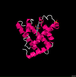
<Structure load='1mbo' size='500' frame='true' align='right' caption='oxymyoglobin ([[1mbo]])' scene='User:Michael_Patrick/Sandbox_1/1mbo-2/1' /> | <Structure load='1mbo' size='500' frame='true' align='right' caption='oxymyoglobin ([[1mbo]])' scene='User:Michael_Patrick/Sandbox_1/1mbo-2/1' /> | ||
| - | This representation shows the protein as blue strands surround the heme ligand, with accompanying water molecules. This water is strongly attracted to the protein and is part of the structure of any crystalline protein. <scene name='User:Michael_Patrick/Sandbox_1/1mbo- | + | This representation shows the protein as blue strands surround the heme ligand, with accompanying water molecules. This water is strongly attracted to the protein and is part of the structure of any crystalline protein. <scene name='User:Michael_Patrick/Sandbox_1/1mbo-4/1'>Hiding the water</scene>reveals that the overall tertiary shape is much like a hockey puck. The α-helix is a prominent secondary structural component. The α-helices can be shown to form two layers of backbone, and myoglobin can be classified as an antiparallel α-helix type of globular protein. The [[Myoglobin]] page gives more detail on the secondary structure. The Ramachandran plot shows most of the residues involved in an α-helix are clustered in the area of the plot where one would expect them to be. (Review Ramachandran Plot.) Many of the residues that are outside of the expected cluster are at the end of a helix, and it is not unusual for such residues to have ψ and φ values that are outside of the range for the α-helix. Also notice that many of the residues that are in the quadrants on the right are Gly. (Residues can be identified by hovering over the sphere with the cursor.) The prosthetic group of myoglobin is a heme, and as shown here it is inserted into a pocket which is nonpolar. Empty heme pocket lined with translucent surface shows that except for some oxygen on the bottom and His 93 at the mid point of one side the pocket is lined with nonpolar carbon atoms. The mostly nonpolar heme inserts into this pocket with the two carboxylate groups of the heme being on the molecular surface. Detailed description of heme structure. The heme shown in the pocket with the pocket's surface colored white so that the heme can be distinguished from the protein surface atoms. His 93 is the fifth ligand chelated to Fe2+ (the other four are the nitrogens in the pyrole rings), and it binds to one side of the heme. Show protein atoms displayed as spacefill that are within 0.5 nm of the heme. These are the atoms which form the surface of the heme pocket and serve as a reminder that except for the ones on the surface of the molecule most of these atoms are carbon atoms and produce a nonpolar environment for the heme. This nonpolar, water-excluding environment is important for the function of myoglobin. Whenever Fe2+ is in an aqueous environment and it contacts O2, Fe2+ is oxidized to Fe3+. Myoglobin with a heme containing Fe3+ (called metmyoglobin) can not fulfill its physiological function and therefore must be degraded |
Revision as of 20:08, 4 March 2011
Contents |
MYOGLOBIN
Background
Myoglobin is an iron- and oxygen-binding protein found in the muscle tissue of vertebrates in general and in almost all mammals. It is related to hemoglobin, which is the iron- and oxygen-binding protein in blood, specifically in the red blood cells. The only time myoglobin is found in the bloodstream is when it is released following muscle injury. It is an abnormal finding, and can be diagnostically relevant when found in blood.
Myoglobin (abbreviated Mb) is a single-chain globular protein of 153 or 154 amino acids, containing a heme (iron-containing porphyrin) prosthetic group in the center around which the remaining apoprotein folds. It has eight alpha helices and a hydrophobic core. It has a molecular weight of 16,700 daltons, and is the primary oxygen-carrying pigment of muscle tissues. Unlike the blood-borne hemoglobin, to which it is structurally related, this protein does not exhibit cooperative binding of oxygen, since positive cooperativity is a property of multimeric/oligomeric proteins only. Instead, the binding of oxygen by myoglobin is unaffected by the oxygen pressure in the surrounding tissue. Myoglobin is often cited as having an "instant binding tenacity" to oxygen given its hyperbolic oxygen dissociation curve. High concentrations of myoglobin in muscle cells allow organisms to hold their breaths longer. Diving mammals such as whales and seals have muscles with particularly high myoglobin abundance.
Myoglobin was the first protein to have its three-dimensional structure revealed. In 1958, John Kendrew and associates successfully determined the structure of myoglobin by high-resolution X-ray crystallography. For this discovery, John Kendrew shared the 1962 Nobel Prize in chemistry with Max Perutz. Despite being one of the most studied proteins in biology, its true physiological function is not yet conclusively established: mice genetically engineered to lack myoglobin are viable, but showed a 30% reduction in cardiac systolic output. They adapted to this deficiency through hypoxic genetic mechanisms and increased vasodilation. In humans myoglobin is encoded by the MB gene.
PDB Entry
1MBO is a 1 chain structure with sequence from Physeter catodon. The January 2000 RCSB PDB Molecule of the Month feature on Myoglobin by David S. Goodsell is 10.2210/rcsb_pdb/mom_2000_1. Full crystallographic information is available from OCA.
About This Structure
|
This representation shows the protein as blue strands surround the heme ligand, with accompanying water molecules. This water is strongly attracted to the protein and is part of the structure of any crystalline protein. reveals that the overall tertiary shape is much like a hockey puck. The α-helix is a prominent secondary structural component. The α-helices can be shown to form two layers of backbone, and myoglobin can be classified as an antiparallel α-helix type of globular protein. The Myoglobin page gives more detail on the secondary structure. The Ramachandran plot shows most of the residues involved in an α-helix are clustered in the area of the plot where one would expect them to be. (Review Ramachandran Plot.) Many of the residues that are outside of the expected cluster are at the end of a helix, and it is not unusual for such residues to have ψ and φ values that are outside of the range for the α-helix. Also notice that many of the residues that are in the quadrants on the right are Gly. (Residues can be identified by hovering over the sphere with the cursor.) The prosthetic group of myoglobin is a heme, and as shown here it is inserted into a pocket which is nonpolar. Empty heme pocket lined with translucent surface shows that except for some oxygen on the bottom and His 93 at the mid point of one side the pocket is lined with nonpolar carbon atoms. The mostly nonpolar heme inserts into this pocket with the two carboxylate groups of the heme being on the molecular surface. Detailed description of heme structure. The heme shown in the pocket with the pocket's surface colored white so that the heme can be distinguished from the protein surface atoms. His 93 is the fifth ligand chelated to Fe2+ (the other four are the nitrogens in the pyrole rings), and it binds to one side of the heme. Show protein atoms displayed as spacefill that are within 0.5 nm of the heme. These are the atoms which form the surface of the heme pocket and serve as a reminder that except for the ones on the surface of the molecule most of these atoms are carbon atoms and produce a nonpolar environment for the heme. This nonpolar, water-excluding environment is important for the function of myoglobin. Whenever Fe2+ is in an aqueous environment and it contacts O2, Fe2+ is oxidized to Fe3+. Myoglobin with a heme containing Fe3+ (called metmyoglobin) can not fulfill its physiological function and therefore must be degraded
Role in Disease
Myoglobin is released from damaged muscle tissue (rhabdomyolysis), which has very high concentrations of myoglobin. The released myoglobin is filtered by the kidneys but is toxic to the renal tubular epithelium and so may cause acute renal failure.
Myoglobin is a sensitive marker for muscle injury, making it a potential marker for heart attack in patients with chest pain. However, elevated myoglobin has low specificity for acute myocardial infarction (AMI) and thus CK-MB, cTnT, ECG, and clinical signs should be taken into account to make the diagnosis.
Reference for the Structure
- Phillips SE. Structure and refinement of oxymyoglobin at 1.6 A resolution. J Mol Biol. 1980 Oct 5;142(4):531-54. PMID:7463482

