Potassium Channel
From Proteopedia
| Line 12: | Line 12: | ||
====Voltage Sensor==== | ====Voltage Sensor==== | ||
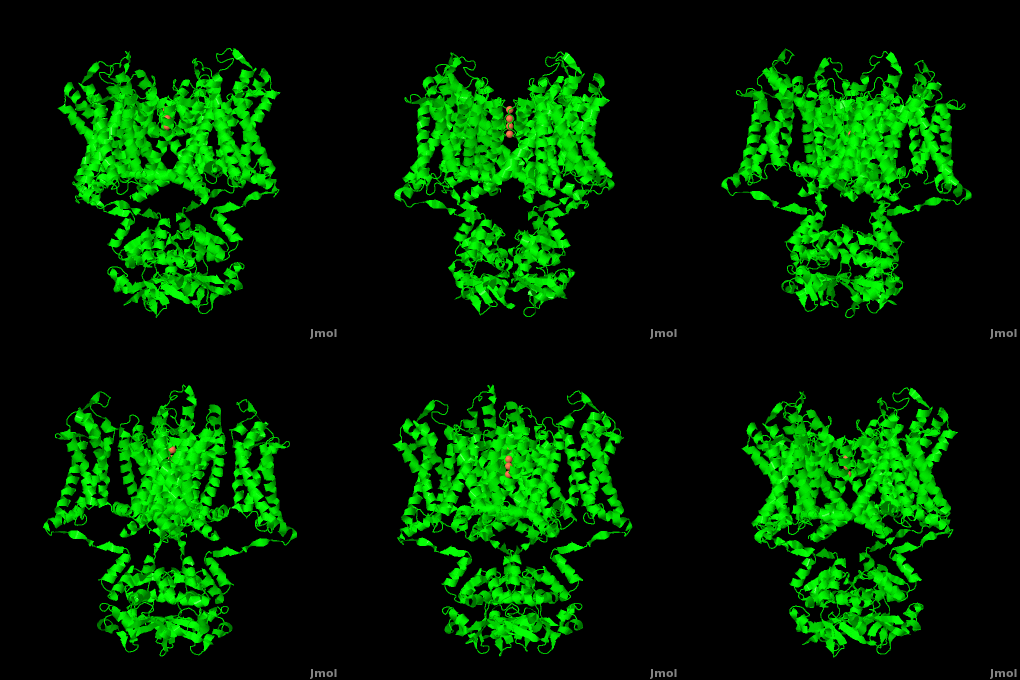
| - | Channel pore opening is dependent on the membrane voltage, a characteristic that is “sensed” by the <scene name='Potassium_Channel/Voltage_sensors_opening/6'>voltage sensor</scene>. The voltage sensor is comprised of <scene name='Potassium_Channel/Voltage_helices/3'>six helices</scene>, S0, S1, S2, S3, S4, & S5. Negatively charged amino acids in the sensor are either located in the <scene name='Potassium_Channel/Voltage_external/3'>external cluster</scene>, consisting of Glu 183 (in the [[2r9r]] structure) and Glu 226, or in the <scene name='Potassium_Channel/Voltage_internal/4'>internal cluster</scene> consisting of Glu 154, Glu 236, and Asp 259. The external cluster is exposed to solvent while the internal cluster is buried. <scene name='Potassium_Channel/Voltage_phe/2'>Phenylalanine 233</scene> separates the external and internal clusters.<ref name="Long"/> The 7 <scene name='Potassium_Channel/Voltage_phe/3'>positively charged residues</scene> of the voltage sensor are located on the S4 helix. Lys 302 and Arg 305 <scene name='Potassium_Channel/Voltage_lower_pos/1'>form hydrogen bonds</scene> with the internal negative cluster while Arginines 287, 290, 293, 296 and 299 are <scene name='Potassium_Channel/Voltage_solvent/1'>exposed to the extracellular solution</scene> (<scene name='Potassium_Channel/Voltage_sensors_arginines_over/2'>Overview</scene>). When the voltage sensor is exposed to a strong negative electric field in the intracellular membrane, the positive gating charges shift inward with the α-carbon of Arg 290 shifting to interact with Phe 233. This shift effectively squeezes the pore shut, closing the intracellular-extracellular pathway. For a comparison see: The <scene name='Potassium_Channel/Open/1'>Open</scene> Channel vs. The <scene name='Potassium_Channel/Closed/1'>Closed</scene> ([[1k4c]]) Channel.<ref name="Long"/> Or view the | + | Channel pore opening is dependent on the membrane voltage, a characteristic that is “sensed” by the <scene name='Potassium_Channel/Voltage_sensors_opening/6'>voltage sensor</scene>. The voltage sensor is comprised of <scene name='Potassium_Channel/Voltage_helices/3'>six helices</scene>, S0, S1, S2, S3, S4, & S5. Negatively charged amino acids in the sensor are either located in the <scene name='Potassium_Channel/Voltage_external/3'>external cluster</scene>, consisting of Glu 183 (in the [[2r9r]] structure) and Glu 226, or in the <scene name='Potassium_Channel/Voltage_internal/4'>internal cluster</scene> consisting of Glu 154, Glu 236, and Asp 259. The external cluster is exposed to solvent while the internal cluster is buried. <scene name='Potassium_Channel/Voltage_phe/2'>Phenylalanine 233</scene> separates the external and internal clusters.<ref name="Long"/> The 7 <scene name='Potassium_Channel/Voltage_phe/3'>positively charged residues</scene> of the voltage sensor are located on the S4 helix. Lys 302 and Arg 305 <scene name='Potassium_Channel/Voltage_lower_pos/1'>form hydrogen bonds</scene> with the internal negative cluster while Arginines 287, 290, 293, 296 and 299 are <scene name='Potassium_Channel/Voltage_solvent/1'>exposed to the extracellular solution</scene> (<scene name='Potassium_Channel/Voltage_sensors_arginines_over/2'>Overview</scene>). When the voltage sensor is exposed to a strong negative electric field in the intracellular membrane, the positive gating charges shift inward with the α-carbon of Arg 290 shifting to interact with Phe 233. This shift effectively squeezes the pore shut, closing the intracellular-extracellular pathway. For a comparison see: The <scene name='Potassium_Channel/Open/1'>Open</scene> Channel vs. The <scene name='Potassium_Channel/Closed/1'>Closed</scene> ([[1k4c]]) Channel.<ref name="Long"/> Or view the morph of the <scene name='Potassium_Channel/Morph/3'>channel opening and closing</scene> (<scene name='Potassium_Channel/Morph/4'>Cartoon Design</scene>). |
Overall, Potassium channels are remarkable structures that allow for near diffusion limit transfer of molecules with sub-angstrom specificity. Our understanding of the structure of Potassium Channels has opened up the potential for [[Pharmaceutical drugs|therapeutic intervention]] into Potassium channel related diseases. | Overall, Potassium channels are remarkable structures that allow for near diffusion limit transfer of molecules with sub-angstrom specificity. Our understanding of the structure of Potassium Channels has opened up the potential for [[Pharmaceutical drugs|therapeutic intervention]] into Potassium channel related diseases. | ||
Revision as of 03:47, 9 March 2011
| |||||||||||
Additional Structures of Potassium Channels
2k44 – KCh voltage-sensor paddle domain – NMR
3e86 – BcKCh transmembrane domain – Bacillus cereus
3e8g – BcKCh transmembrane domain +Ca+Na
2q67, 2q68, 2q69, 2q6a – BcKCh (mutant)
2ahy – BcKCh+Na
2ahz – BcKCh+K
2pnv – rKCh leucine zipper domain SKCa – rat
2nz0 – hKCh Kv4.3 N-terminal+KV channel interacting protein 1 – human
2i2r - rKCh Kv4.3 N-terminal+KV channel interacting protein 1
1s6c - rKCh Kv4.2 N-terminal+KV channel interacting protein 1
1qx7 – rKCh SKCa+ calmodulin
1g4y – rKCh rSK2 calmodulin binding domain SKCa + calmodulin
1j95 – KCh+K+tetrabutylammonium – Streptomyces lividans
3co2, 1vp6 – MlotiK1 cyclic nucleotide binding domain – Mesorhizobium loti
For Additional Structures, See: Potassium Channels
Additional Resources
For Additional Information, See: Membrane Channels & Pumps
References
- ↑ 1.0 1.1 Zhou Y, Morais-Cabral JH, Kaufman A, MacKinnon R. Chemistry of ion coordination and hydration revealed by a K+ channel-Fab complex at 2.0 A resolution. Nature. 2001 Nov 1;414(6859):43-8. PMID:11689936 doi:http://dx.doi.org/10.1038/35102009
- ↑ 2.0 2.1 2.2 2.3 Doyle DA, Morais Cabral J, Pfuetzner RA, Kuo A, Gulbis JM, Cohen SL, Chait BT, MacKinnon R. The structure of the potassium channel: molecular basis of K+ conduction and selectivity. Science. 1998 Apr 3;280(5360):69-77. PMID:9525859
- ↑ Jiang Y, Lee A, Chen J, Ruta V, Cadene M, Chait BT, MacKinnon R. X-ray structure of a voltage-dependent K+ channel. Nature. 2003 May 1;423(6935):33-41. PMID:12721618 doi:http://dx.doi.org/10.1038/nature01580
- ↑ Waters MF, Minassian NA, Stevanin G, Figueroa KP, Bannister JP, Nolte D, Mock AF, Evidente VG, Fee DB, Muller U, Durr A, Brice A, Papazian DM, Pulst SM. Mutations in voltage-gated potassium channel KCNC3 cause degenerative and developmental central nervous system phenotypes. Nat Genet. 2006 Apr;38(4):447-51. Epub 2006 Feb 26. PMID:16501573 doi:ng1758
- ↑ 5.0 5.1 5.2 5.3 Long SB, Tao X, Campbell EB, MacKinnon R. Atomic structure of a voltage-dependent K+ channel in a lipid membrane-like environment. Nature. 2007 Nov 15;450(7168):376-82. PMID:18004376 doi:http://dx.doi.org/10.1038/nature06265
Proteopedia Page Contributors and Editors (what is this?)
Michal Harel, David Canner, Joel L. Sussman, Alexander Berchansky, Ann Taylor


