Sandbox Reserved 350
From Proteopedia
| Line 40: | Line 40: | ||
*'''Apex 3'''—<font color='deeppink'>'''Gly75-Tyr84;''' Hydrophobic Loop (Leu79). </font> | *'''Apex 3'''—<font color='deeppink'>'''Gly75-Tyr84;''' Hydrophobic Loop (Leu79). </font> | ||
| - | <br /> | ||
| - | |||
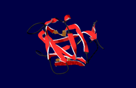
<Structure load='1czv' size='' frame='true' align='right' caption=' C2 Domain of Human Coagulation Factor V' scene='Sandbox_Reserved_350/Expriment3/2'>TextToBeDisplayed</scene>'/> | <Structure load='1czv' size='' frame='true' align='right' caption=' C2 Domain of Human Coagulation Factor V' scene='Sandbox_Reserved_350/Expriment3/2'>TextToBeDisplayed</scene>'/> | ||
| - | + | ||
| - | + | The apexes of these <font color='orangered'> '''three loops''' </font> within the C2 domain, are able to create a deep groove lined by '''hydrophobic''' <font color='brown'> '''(Trp31, Met83)''' </font> and '''polar residues''' <font color='royalblue'> '''(Gln48, Ser78)'''</font>, as seen and consisting the <scene name='Sandbox_Reserved_350/Expriment3/3'> "Open Form" </scene> of FVa-C2. This groove is seen as the primary membrane-binding site of the C2-Domain. <ref name="Pubmed"/> | |
| - | The apexes of these <font color='orangered'> '''three loops''' </font> within the C2 domain, are able to create a deep groove lined by '''hydrophobic''' <font color='brown'> '''(Trp31, Met83)''' </font> and '''polar residues''' <font color='royalblue'> '''(Gln48, Ser78)'''</font>, as seen and consisting the '' | + | |
<br /> | <br /> | ||
A second dimeric crystal form of FVa-C2, packed through the free edges of S6 strands, presenting a different Leu104-Val109 | A second dimeric crystal form of FVa-C2, packed through the free edges of S6 strands, presenting a different Leu104-Val109 | ||
| Line 55: | Line 52: | ||
The three loops are described by ''Macedo-Ribeiro et al.'' to protrude like spikes from the bottom of the barrel in monomeric FVa-C2.<ref name="Pubmed"/> It is also worth noting that spike (1) & spike (3) are separated by β-hairpin structures and spike (2) is described as a wider irregularly loop comparatively. These three loops extending from the C2 domain, are all linked to each other, and to '''three shorter loops''' by an intricate '''H-bonding network''' which extends to residues at the bottom of the β-barrel.<ref name="Pubmed"/> | The three loops are described by ''Macedo-Ribeiro et al.'' to protrude like spikes from the bottom of the barrel in monomeric FVa-C2.<ref name="Pubmed"/> It is also worth noting that spike (1) & spike (3) are separated by β-hairpin structures and spike (2) is described as a wider irregularly loop comparatively. These three loops extending from the C2 domain, are all linked to each other, and to '''three shorter loops''' by an intricate '''H-bonding network''' which extends to residues at the bottom of the β-barrel.<ref name="Pubmed"/> | ||
<br /> | <br /> | ||
| - | The overall Barrel structure is closed at the top and bottom by | + | The overall Barrel structure is closed at the top and bottom by straight segments, giving it an overall spherical shape with a flattened upper surface. |
<br /> | <br /> | ||
Revision as of 02:16, 4 April 2011
| This Sandbox is Reserved from January 10, 2010, through April 10, 2011 for use in BCMB 307-Proteins course taught by Andrea Gorrell at the University of Northern British Columbia, Prince George, BC, Canada. |
To get started:
More help: Help:Editing |
- Protein: cHuman Coagulation factor V, 1czv [1]
Introduction
Coagulation Factor V studied in 1987 by William H. Kane, Akitada Ichinose, Frederick S. Hagen and Earl W. Davie, out of University of Washington, Seattle [2]
| |||||||||
| 1czv, resolution 2.40Å () | |||||||||
|---|---|---|---|---|---|---|---|---|---|
| Related: | 1czs, 1czt | ||||||||
| |||||||||
| |||||||||
| Resources: | FirstGlance, OCA, RCSB, PDBsum | ||||||||
| Coordinates: | save as pdb, mmCIF, xml | ||||||||
Contents |
Structure & Function
The structure of Human Coagulation Factor V (FV) precursors from a translated polypeptide
to a A1-A2-B-A3-C1-C2 layout which results in the activated (FVa) protein.[1]
- Heavy A1-A2 Chain
- Light A3-C1-C2 Chain
FVa consists of a conserved β-Barrel framework acting as a scaffold for three loops of the C2 domain (FVa-C2).[1]
The FVa-C2, which is classified as a distorted jelly-roll , which is compossed of arranged into two β-sheets of five and three strands packed against one another.
Salt bridges located within the "upper" segment (Asp61-Arg134) . The C2-Domain of Human coagulation factor is homologous to a larger family of adhesion proteins; Discoidin,but not related to synaptotagmin-like C2 domains.[1]
[1]
- Apex 1—Ser21-Trp31; containing Indole moieties able to form hydrogen bonds (Involving two consecutive Trp 26 & 27).
- Apex 2—Asn39-Asn45; capped with a basic residue able to form hydrogen bonds (Arg43).
- Apex 3—Gly75-Tyr84; Hydrophobic Loop (Leu79).
| |||||||||||


