Sandbox Reserved 312
From Proteopedia
| Line 14: | Line 14: | ||
The HRDC domain has 80 amino acids at the C-terminal end. There are five helicases connected by turns and hydrophobic loops. These hydrobopic loops are needed for for packing the domain into a bundle. This domain contributes in the interaction with the double stranded DNA. | The HRDC domain has 80 amino acids at the C-terminal end. There are five helicases connected by turns and hydrophobic loops. These hydrobopic loops are needed for for packing the domain into a bundle. This domain contributes in the interaction with the double stranded DNA. | ||
| - | The <scene name='Sandbox_Reserved_312/Active_site/1'>active site</scene> catalysis the exonuclease activity by having the <scene name='Sandbox_Reserved_312/two metal ions/2'>two metal ions</scene> Mn<sup>2+</sup> or Mg<sup>2+</sup> complex. Mg<sup>2+</sup> are more commonly cofactors in the cell; however, Mn<sup>2+</sup> supports higher catalytic activity. This active site contains Glu<sub>84</sub>, Tyr<sub>212</sub>, Ala<sub>188</sub>, Glu<sub>192</sub>, Asp<sub>216</sub>, Asp<sub>82</sub>, Tyr<sub>54</sub> and Asp<sub>143</sub>. The two metals have a distance of 3.7 Å separating them when bound to the active site. The inner Mn<sup>2+</sup> is directly coordinated with Asp<sub>82</sub>, Glu<sub>84</sub> and Asp<sub>216</sub>. The outer metal ion binds with a ligatures side chain Asp<sub>82</sub> to form the bridge between the two metal ions. | + | The <scene name='Sandbox_Reserved_312/Active_site/1'>active site</scene> catalysis the exonuclease activity by having the <scene name='Sandbox_Reserved_312/two metal ions/2'>two metal ions</scene> Mn<sup>2+</sup> or Mg<sup>2+</sup> complex. Mg<sup>2+</sup> are more commonly cofactors in the cell; however, Mn<sup>2+</sup> supports higher catalytic activity. This active site contains Glu<sub>84</sub>, Tyr<sub>212</sub>, Ala<sub>188</sub>, Glu<sub>192</sub>, Asp<sub>216</sub>, Asp<sub>82</sub>, Tyr<sub>54</sub> and Asp<sub>143</sub>.<ref name= "Perry"> PMID: 16622405 </ref> The two metals have a distance of 3.7 Å separating them when bound to the active site.<ref name= "Perry"> PMID: 16622405 </ref> The inner Mn<sup>2+</sup> is directly coordinated with Asp<sub>82</sub>, Glu<sub>84</sub> and Asp<sub>216</sub>.<ref name= "Perry"> PMID: 16622405 </ref> The outer metal ion binds with a ligatures side chain Asp<sub>82</sub> to form the bridge between the two metal ions.<ref name= "Perry"> PMID: 16622405 </ref> Asp<sub>143</sub> interacts indirectly with the outer metal ion by two water molecules. <ref name= "Perry"> PMID: 16622405 The binding of the two metal ions do not change conformation of the WRN protein but the correct bond is vital for the activity of the WRN exonuclease.<ref name= "Perry"> PMID: 16622405 |
The protein has two conserved ATP binding sites with a location for Mn cations association. This binding and hydolysis is depenent on Mn<sup>2+</sup> binding. | The protein has two conserved ATP binding sites with a location for Mn cations association. This binding and hydolysis is depenent on Mn<sup>2+</sup> binding. | ||
Revision as of 03:03, 4 April 2011
| This Sandbox is Reserved from January 10, 2010, through April 10, 2011 for use in BCMB 307-Proteins course taught by Andrea Gorrell at the University of Northern British Columbia, Prince George, BC, Canada. |
To get started:
More help: Help:Editing |
Contents |
WRN exonuclease
| |||||||||
| 2fbv, resolution 2.40Å () | |||||||||
|---|---|---|---|---|---|---|---|---|---|
| Ligands: | |||||||||
| Gene: | WRN, RECQ3, RECQL2 (Homo sapiens) | ||||||||
| Related: | 2fbt, 2fbx, 2fby, 2fc0 | ||||||||
| |||||||||
| |||||||||
| Resources: | FirstGlance, OCA, RCSB, PDBsum | ||||||||
| Coordinates: | save as pdb, mmCIF, xml | ||||||||
Introduction
Werner (WRN) protein is a member of the human RecQDNA helicases. This enzyme has a helicases function (unwinding double-stranded DNA helix in the 3'-5' direction) and a exonuclease activity (degrading), which allows for deletion of mutation and proofreading. The WRN has a functional exonuclease domain located on the N-terminus. WRN protein protects human from cancer; as well as, premature aging.
Structure
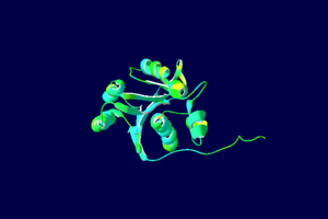
The WRN protein is built from several individual WRN copies. The exonuclease region of the protein is located at the N-terminal end with 171 amino acids. They form a hexamer around the end of the strand when exposed to double strand DNA, which is essential to DNA repair of the WRN.
The HRDC domain has 80 amino acids at the C-terminal end. There are five helicases connected by turns and hydrophobic loops. These hydrobopic loops are needed for for packing the domain into a bundle. This domain contributes in the interaction with the double stranded DNA.
The catalysis the exonuclease activity by having the Mn2+ or Mg2+ complex. Mg2+ are more commonly cofactors in the cell; however, Mn2+ supports higher catalytic activity. This active site contains Glu84, Tyr212, Ala188, Glu192, Asp216, Asp82, Tyr54 and Asp143.[1] The two metals have a distance of 3.7 Å separating them when bound to the active site.[1] The inner Mn2+ is directly coordinated with Asp82, Glu84 and Asp216.[1] The outer metal ion binds with a ligatures side chain Asp82 to form the bridge between the two metal ions.[1] Asp143 interacts indirectly with the outer metal ion by two water molecules. [1] This provides energy for DNA helicase, breaking hydrogen bonds between bases of double stranded DNA, which unwinds the helix.
A six membered WRN ring is formed to fit around the DNA helix. The exonuclease active site and Mn2+ point inward towards the DNA.
Function
This protein plays a major role in DNA metabolic pathway, which perform DNA repair and genome stability. The protein is a 3`-5`helicase which indicates that it can not be used to transcription and replication (5`-3`). WRN has activity of ATPase, helicases and exonuclease. [2] WRN exonuclease functions on different structures of a DNA substrate.[1] The metal ions Mn2+ are needed to stimulate the exonuclease active of the WRN. However, WRN exonuclease can be blocked by oxidatively induced base lesion (damaged by disease tissue). The homodimer protein Ku binds to the broken double-stranded DNA, stimulating the WRN exonuclease to bypass the lesion. However, in non-digested strands the process is blocked by the lesion.
WRN are found in tissues, mainly in the pancreas and testis. In cells, WRN is found in the nucleus and nucleolar regions.
Clinical significance
The build up of oxidatively induced base in DNA the gene coding for WRN is mutated a autosomal recessive disorder causing rapid aging(osteoporosis, atherosclerosis and cancer). This is known as Werner syndrome which appears at puberty.
Mutation in the WRN gene occurs int the HRDC domain. This domain actives the DNA association with the helicase; as a result, a mutation in this region blocks DNA repair mechanism to occur. As DNA cell damage accumulates cell death occurs losing WRN protein function, leading to premature aging
Reference
- ↑ 1.0 1.1 1.2 1.3 1.4 1.5 Perry JJ, Yannone SM, Holden LG, Hitomi C, Asaithamby A, Han S, Cooper PK, Chen DJ, Tainer JA. WRN exonuclease structure and molecular mechanism imply an editing role in DNA end processing. Nat Struct Mol Biol. 2006 May;13(5):414-22. Epub 2006 Apr 23. PMID:16622405 doi:http://dx.doi.org/10.1038/nsmb1088
- ↑ Perry JJ, Yannone SM, Holden LG, Hitomi C, Asaithamby A, Han S, Cooper PK, Chen DJ, Tainer JA. WRN exonuclease structure and molecular mechanism imply an editing role in DNA end processing. Nat Struct Mol Biol. 2006 May;13(5):414-22. Epub 2006 Apr 23. PMID:16622405 doi:http://dx.doi.org/10.1038/nsmb1088
<references/"Bukowy"/WRN Exonuclease activity is blocked by specific oxidatively induced base lesions positioned in either DNA strand>


