User:R. Jeremy Johnson/RNaseS RNaseB
From Proteopedia
| Line 9: | Line 9: | ||
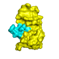
[[Image:surface view of rnase s.png|200px|left|thumb| The image above depicts the surface interaction between the S peptide and S protein fragments; S peptide is blue, S protein is yellow.]] | [[Image:surface view of rnase s.png|200px|left|thumb| The image above depicts the surface interaction between the S peptide and S protein fragments; S peptide is blue, S protein is yellow.]] | ||
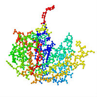
| - | + | <Structure load='1RNU' size='250' frame='true' align='right' caption='RNase S (PDB: 1RNU) is a protein composed of 2 fragments: S Peptide (residues 1-20) and S Protein (residues 21-124). It is a result of cleavage of RNase A between residues 20 and 21.' scene='Sandbox_Reserved_195/Ribonuclese_s_basic/3' target='0' /> | |
| - | + | ||
| - | <Structure load='1RNU' size=' | + | |
| + | RNase A and RNase S have very similar structures except for a few key areas, one being the cleavage site (residues 16-23)<ref name="Raines"> PMID:11848924</ref>. Decreased order in these amino acids is seen in RNase S. The <scene name='Sandbox_Reserved_195/Clevage_site/4' target='0'>cleavage site</scene> on RNase A is located between residues Ala 20 and Ser 21. Upon cleavage, the fragments S peptide and S protein are created, giving RNase S its unique structure. <scene name='Sandbox_Reserved_195/S_peptide/8' target='0'>S Peptide</scene> consists of residues 1-20, and is largely responsible for proper folding. <scene name='Sandbox_Reserved_195/S_protein/2'>S protein</scene> is comprised of residues 21-124. Additionally, RNase A and RNase S have many conserved structural components, as they are essentially the same protein. Glutamine 60 is highly conserved structurally between <scene name='Sandbox_Reserved_195/Gln_60/3' target='0'>RNase S</scene> and <scene name='Sandbox_Reserved_195/Glutamine_60_rnase_a/5' target='0'>RNase A</scene>, as well as in many different species, demonstrating its importance. Glu 60 is also interesting, because it is the only residue in an unfavorable position (Φ= -100, ψ= -130) as defined in the Ramachandran plot <ref name="Kim">PMID:1463719</ref>. | ||
The <scene name='Sandbox_Reserved_195/Active_site/2' target='0'>active site</scene> of RNase S consists of <scene name='Sandbox_Reserved_195/Active_site/3'>residues</scene> His 12, Lys 41, Val 43, Asn 44, Thr 45, His 119, Phe 120, Asp 121, and Ser 123. <scene name='Sandbox_Reserved_195/His_12/1'>His 12</scene> is the only residue in the active site that is located on the S peptide fragment. <scene name='Sandbox_Reserved_195/Lys_41_and_his_119/1'>Lys 41 and His 119</scene> are also crucial to the functioning of RNase S. Interestingly, both catalytic Histidines perform the same function, even though they are located on <scene name='Sandbox_Reserved_195/Histines_and_lysines/2'>different fragments</scene>. Residues 1, 15-20, 21-23, and 124 could be removed without serious consequence to the structure or activity of RNase S. Residue 124 could also be removed from RNase A without compromising the structure. <ref name="Wyckoff et al">PMID:6037556</ref> | The <scene name='Sandbox_Reserved_195/Active_site/2' target='0'>active site</scene> of RNase S consists of <scene name='Sandbox_Reserved_195/Active_site/3'>residues</scene> His 12, Lys 41, Val 43, Asn 44, Thr 45, His 119, Phe 120, Asp 121, and Ser 123. <scene name='Sandbox_Reserved_195/His_12/1'>His 12</scene> is the only residue in the active site that is located on the S peptide fragment. <scene name='Sandbox_Reserved_195/Lys_41_and_his_119/1'>Lys 41 and His 119</scene> are also crucial to the functioning of RNase S. Interestingly, both catalytic Histidines perform the same function, even though they are located on <scene name='Sandbox_Reserved_195/Histines_and_lysines/2'>different fragments</scene>. Residues 1, 15-20, 21-23, and 124 could be removed without serious consequence to the structure or activity of RNase S. Residue 124 could also be removed from RNase A without compromising the structure. <ref name="Wyckoff et al">PMID:6037556</ref> | ||
| Line 19: | Line 18: | ||
== Introduction to Ribonuclease B == | == Introduction to Ribonuclease B == | ||
| - | |||
| - | <Structure load='1rbb' size='300' frame='true' align='right' caption='3D picture of RNase B dimer' scene='Sandbox_Reserved_196/Rbb_basic/1' name='1rbb' /> | ||
Ribonuclease B (RNase B) is a form of the enzyme RNase A that has an added glycoprotein with N-linked carbohydrates at <scene name='Sandbox_Reserved_196/Rbb_basic/16'>Asn-34</scene>, which means that the carbohydrate is attached at the <scene name='Sandbox_Reserved_196/Rbb_basic/17'>nitrogen</scene> of the Aspargine-34 side chain. This added sugar chain can aid in the folding of the protein as well as in cell to cell signaling<ref name="first">Varki A, Cummings RD, Esko JD, Freeze HH, Stanley P, Bertozzi CR, Hart GW, Etzler ME. ''Essentials of Glycobiology''. PMID:20301239</ref>. Glycoproteins also play an important role in tumor formation because it has been found that N-linked glycans recognized by the CD337 receptor on "natural killer cells" are mutated in tumor cells, stopping their death<ref name="second">http://en.wikipedia.org/wiki/Glycan</ref>. Other than the attachment of the polysaccharide, RNase B is structurally the same as RNase A, but the attachment allows for additional catalytic activity. This small change allows RNase B to hydrolyze double-stranded RNA at ionic strengths where RNase A has no activity, showing that small changes in the active sites of very similar molecules can lead to new roles and activities <ref name="third">PMID:3680242</ref>. <scene name='Sandbox_Reserved_196/Rbb_basic/1'>(Return to original scene)</scene> | Ribonuclease B (RNase B) is a form of the enzyme RNase A that has an added glycoprotein with N-linked carbohydrates at <scene name='Sandbox_Reserved_196/Rbb_basic/16'>Asn-34</scene>, which means that the carbohydrate is attached at the <scene name='Sandbox_Reserved_196/Rbb_basic/17'>nitrogen</scene> of the Aspargine-34 side chain. This added sugar chain can aid in the folding of the protein as well as in cell to cell signaling<ref name="first">Varki A, Cummings RD, Esko JD, Freeze HH, Stanley P, Bertozzi CR, Hart GW, Etzler ME. ''Essentials of Glycobiology''. PMID:20301239</ref>. Glycoproteins also play an important role in tumor formation because it has been found that N-linked glycans recognized by the CD337 receptor on "natural killer cells" are mutated in tumor cells, stopping their death<ref name="second">http://en.wikipedia.org/wiki/Glycan</ref>. Other than the attachment of the polysaccharide, RNase B is structurally the same as RNase A, but the attachment allows for additional catalytic activity. This small change allows RNase B to hydrolyze double-stranded RNA at ionic strengths where RNase A has no activity, showing that small changes in the active sites of very similar molecules can lead to new roles and activities <ref name="third">PMID:3680242</ref>. <scene name='Sandbox_Reserved_196/Rbb_basic/1'>(Return to original scene)</scene> | ||
| Line 26: | Line 23: | ||
== Structure and Biology of RNase B == | == Structure and Biology of RNase B == | ||
[[Image:1RBJ.jpg |200px|left|thumb|RNaseB]] | [[Image:1RBJ.jpg |200px|left|thumb|RNaseB]] | ||
| + | |||
| + | <Structure load='1rbb' size='300' frame='true' align='right' caption='3D picture of RNase B dimer' scene='Sandbox_Reserved_196/Rbb_basic/1' name='1rbb' /> | ||
Crystallization of RNase A and RNase B has shown that these two enzymes are identical in their<Structure load='1rbj' size='300' frame='true' align='right' caption='Molecule II of Ribonuclease B with a strand of DNA in active site' scene='Sandbox_Reserved_196/Secondary_structure/13' name ='1rbj' /> primary structure and amino acid makeup; however, RNase B has a single glycosylation at the <scene name='Sandbox_Reserved_196/Rbb_basic/18'>Asn-34</scene> site that differentiates RNase B from RNase A. RNase B has from five to nine mannose residues attached which can create a variance within RNase B molecules. This glycosylation increases the kinetic stability of the RNase B by 3 kJ/mol<ref name="fourth">PMID:10600722</ref>. This addition to RNase B, however, does not significantly change the protein conformation from RNase A<ref name="fifth">PMID:1322837</ref>. | Crystallization of RNase A and RNase B has shown that these two enzymes are identical in their<Structure load='1rbj' size='300' frame='true' align='right' caption='Molecule II of Ribonuclease B with a strand of DNA in active site' scene='Sandbox_Reserved_196/Secondary_structure/13' name ='1rbj' /> primary structure and amino acid makeup; however, RNase B has a single glycosylation at the <scene name='Sandbox_Reserved_196/Rbb_basic/18'>Asn-34</scene> site that differentiates RNase B from RNase A. RNase B has from five to nine mannose residues attached which can create a variance within RNase B molecules. This glycosylation increases the kinetic stability of the RNase B by 3 kJ/mol<ref name="fourth">PMID:10600722</ref>. This addition to RNase B, however, does not significantly change the protein conformation from RNase A<ref name="fifth">PMID:1322837</ref>. | ||
| Line 51: | Line 50: | ||
<references group="xtra"/> | <references group="xtra"/> | ||
| + | |||
| + | |||
| + | <Structure load='7rsa' size='300' frame='true' align='right' caption='RNase A (PDB: 7rsa), also known as bovine pancreatic ribonuclease A, has been studied for years to learn about protein structure, stability, and folding.' scene='Sandbox_Reserved_195/Ribonuclease_a_basic/3' target='1' /> | ||
Revision as of 19:17, 7 May 2011
Contents |
Introduction to Ribonuclease Derivatives
Introduction to Ribonuclease S
RNase S is RNase A treated with subtilisin, which cleaves a single peptide bond. Consequently, Ribonuclease S consists of two fragments of bovine Ribonuclease A in a peptide-protein complex: S peptide (amino acids 1-20) and S protein (amino acids 21-124). RNase S was the third enzyme and fourth protein ever cystallized and three-dimensional structure determined. As such, biological work on RNase S helped scientists determine the first 3-dimensional structure of a protein-nucleic acid complex. Additionally, RNase S provided one of the first demonstrations of a crystalline enzyme acting as an active catalyst. Investigations of RNase S have also led to many technological advancements, including substrate leash amplification, fusion protein systems, and protein ubiquitination [1]. RNase S has been studied to reveal mechanisms of protein folding by coupling folding and association. Studies of RNase S has lead to greater interest in the investigation of RNase A and its role in biological systems, and has also lead to a greater knowledge of the correlation between protein folding and enzyme activity.
Structure and Biology of RNase S
|
RNase A and RNase S have very similar structures except for a few key areas, one being the cleavage site (residues 16-23)[1]. Decreased order in these amino acids is seen in RNase S. The on RNase A is located between residues Ala 20 and Ser 21. Upon cleavage, the fragments S peptide and S protein are created, giving RNase S its unique structure. consists of residues 1-20, and is largely responsible for proper folding. is comprised of residues 21-124. Additionally, RNase A and RNase S have many conserved structural components, as they are essentially the same protein. Glutamine 60 is highly conserved structurally between and , as well as in many different species, demonstrating its importance. Glu 60 is also interesting, because it is the only residue in an unfavorable position (Φ= -100, ψ= -130) as defined in the Ramachandran plot [2].
The of RNase S consists of His 12, Lys 41, Val 43, Asn 44, Thr 45, His 119, Phe 120, Asp 121, and Ser 123. is the only residue in the active site that is located on the S peptide fragment. are also crucial to the functioning of RNase S. Interestingly, both catalytic Histidines perform the same function, even though they are located on . Residues 1, 15-20, 21-23, and 124 could be removed without serious consequence to the structure or activity of RNase S. Residue 124 could also be removed from RNase A without compromising the structure. [3]
Hydrogen bonding and hydrophobic interactions play a major role in the structure of RNase S. [2] There are 84 water molecules present, with 8 of these specifically connecting S peptide and S protein. Some of these water molecules are also conserved throughout all RNase derivatives. Hydrophobic interactions between residues Phe 8, Met 13, His 12, Ala 4 are essential in holding the S Peptide in place. In addition to these residues, is also important in peptide-protein binding. In the picture, it appears that the protein and peptide are not connected; this is because the bond cleavage between residues 20 and 21 of RNase A has already occurred. Additionally RNase S has a rigid hydrophobic core; one-third of the surface of the core is made up of the S peptide. The surrounding loops have more flexibility.
Introduction to Ribonuclease B
Ribonuclease B (RNase B) is a form of the enzyme RNase A that has an added glycoprotein with N-linked carbohydrates at , which means that the carbohydrate is attached at the of the Aspargine-34 side chain. This added sugar chain can aid in the folding of the protein as well as in cell to cell signaling[4]. Glycoproteins also play an important role in tumor formation because it has been found that N-linked glycans recognized by the CD337 receptor on "natural killer cells" are mutated in tumor cells, stopping their death[5]. Other than the attachment of the polysaccharide, RNase B is structurally the same as RNase A, but the attachment allows for additional catalytic activity. This small change allows RNase B to hydrolyze double-stranded RNA at ionic strengths where RNase A has no activity, showing that small changes in the active sites of very similar molecules can lead to new roles and activities [6].
Structure and Biology of RNase B
|
|
Slight differences include changes in crystal packing, as the RNase B crystals have two slightly asymmetrical units. The crystals are (shown above) with two separate molecules, and , which are linked by a salt bridge at Asp-121 and Arg-85. This linkage determines the orientation of the two molecules in relation to one another. Not only does a salt bridge link this dimer-type molecule, but other ions also interact via cross-linkage to stabilize the structure [6].
The crystallization of RNase B, in complex with sequence DNA, provided the structure of the active site when bound to nucleic acids. The active site, composed of and found in both molecules I and II of RNase B, is very similar to the active site of RNase A. A difference between RNase A and RNase B is , which are very flexible and can open up or close off the active site. Molecules I and II are slightly asymmetrical, and the most noticeable difference between the two is the position of the . This residue, which is present in both molecules, is much closer to the active site in molecule II. This is important because ions bind to Lys-66, like the DNA, making them accessible to the active site. While the crystalline packing of molecules I and II differ slightly, their active sites bind substrate in the same manner. Even though the crystallization of the structure has been successful, it has not been an aid to providing the mechanism by which RNase B has the catalytic activity to hydrolyze double stranded RNA.[6].
References
- ↑ 1.0 1.1 Raines RT. Ribonuclease A. Chem Rev. 1998 May 7;98(3):1045-1066. PMID:11848924
- ↑ 2.0 2.1 Kim EE, Varadarajan R, Wyckoff HW, Richards FM. Refinement of the crystal structure of ribonuclease S. Comparison with and between the various ribonuclease A structures. Biochemistry. 1992 Dec 15;31(49):12304-14. PMID:1463719
- ↑ Wyckoff HW, Hardman KD, Allewell NM, Inagami T, Johnson LN, Richards FM. The structure of ribonuclease-S at 3.5 A resolution. J Biol Chem. 1967 Sep 10;242(17):3984-8. PMID:6037556
- ↑ Varki A, Cummings RD, Esko JD, Freeze HH, Stanley P, Bertozzi CR, Hart GW, Etzler ME. Essentials of Glycobiology. PMID:20301239
- ↑ http://en.wikipedia.org/wiki/Glycan
- ↑ 6.0 6.1 6.2 Williams RL, Greene SM, McPherson A. The crystal structure of ribonuclease B at 2.5-A resolution. J Biol Chem. 1987 Nov 25;262(33):16020-31. PMID:3680242
- ↑ Imperiali B, O'Connor SE. Effect of N-linked glycosylation on glycopeptide and glycoprotein structure. Curr Opin Chem Biol. 1999 Dec;3(6):643-9. PMID:10600722
- ↑ Joao HC, Scragg IG, Dwek RA. Effects of glycosylation on protein conformation and amide proton exchange rates in RNase B. FEBS Lett. 1992 Aug 3;307(3):343-6. PMID:1322837
Additional Resources
Other resources:
Wikipedia article on RNase A: http://en.wikipedia.org/wiki/RNase_A
RNase S on PDB: http://www.rcsb.org/pdb/explore/explore.do?structureId=1RNU
RNase A on PDB: http://www.rcsb.org/pdb/explore/explore.do?structureId=7RSA
RNase B on PDB: http://www.pdb.org/pdb/explore/explore.do?structureId=1RBJ: http://www.pdb.org/pdb/explore/explore.do?structureId=1RBB
Ribonuclease on Proteopedia: http://proteopedia.org/wiki/index.php/Ribonuclease
|


