Group:MUZIC:Plectin
From Proteopedia

| Line 29: | Line 29: | ||
The plakin domain is formed by an array of spectrin repeats (SR) and a Src-homology 3 (SH3), and harbors binding sites for junctional proteins. This region is adjacent to the actin-binding domain and is required for efficient binding to the integrin alpha6beta4 in hemidesmosomes.<ref>PMID: 21288893 </ref> | The plakin domain is formed by an array of spectrin repeats (SR) and a Src-homology 3 (SH3), and harbors binding sites for junctional proteins. This region is adjacent to the actin-binding domain and is required for efficient binding to the integrin alpha6beta4 in hemidesmosomes.<ref>PMID: 21288893 </ref> | ||
| - | <Structure load='3PE0' size='400' frame='true' align='right' caption='Crystal structure of SR4-SR5-SH3 regions in the plakin domain' scene='User:Jae-Geun_Song/Workbench/plectin/Plakin/1' | + | <Structure load='3PE0' size='400' frame='true' align='right' caption='Crystal structure of SR4-SR5-SH3 regions in the plakin domain' scene='User:Jae-Geun_Song/Workbench/plectin/Plakin/1'> |
Revision as of 13:45, 4 July 2011
|
Contents |
Introduction
Plectin is a multidomain protein with large size (>500kDa) and versatile binding properties, which abundantly expressed in a wide variety of mammalian tissues and cell types, combined with different binding partners. It has important functions in maintaining the mechanical stability of skin, skeletal muscle and heart.
Isoforms
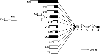
The plectin gene has unusual 5'-end diversity, which is alternatively spliced into exon 2 and makes 11 kinds of isoforms. Expression level of isofroms is varied in tissues,and some of them are specifically expressed in brain,skeletal muscle and skin . [1].
Overall structure
Plectin can be divided in three main sections; a central coiled-coil rod domain, N and C-terminal globular region and exhibits a dumbbell like structure. C-terminal region is composed of 6 homologous repeating domains, and this region has a role in binding to intermediate filaments such as vimentin and cytokeratin [2]. N-terminal globular region contains actin binding domain (ABD) comprising two calponin homology.
Image:Plectin structure.jpg (Litjens, 2006)
Deposited structures in Protein Data Bank(PDB)
Only a few structures are available in PDB and focused on N-terminal region (actin binding domain and plakin domain.
1. Actin Binding Domain (ABD)
Plectin has a canonical actin binding domain in N-terminus, which is consisted of two calponin homology domain()[3]. N-terminal domain of plectin containing ABD interacts with F-actin and regulates actin dynamics in vivo, additionally binding of plectin ABD to vimentin was also reported [4].
2. Plakin domain
The plakin domain is formed by an array of spectrin repeats (SR) and a Src-homology 3 (SH3), and harbors binding sites for junctional proteins. This region is adjacent to the actin-binding domain and is required for efficient binding to the integrin alpha6beta4 in hemidesmosomes.[5]
| |||||||||||
Proteopedia Page Contributors and Editors (what is this?)
Jae-Geun Song, Alexander Berchansky, Nikos Pinotsis, Jaime Prilusky, Michal Harel

