Journal:Cell:1
From Proteopedia
(Difference between revisions)

| Line 23: | Line 23: | ||
====Implications for the binding mode of IFNb==== | ====Implications for the binding mode of IFNb==== | ||
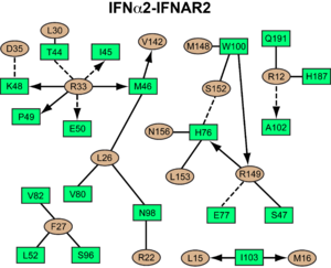
<scene name='User:David_Canner/Workbench2/Ifnbeta/1'>IFNb exhibits</scene> 30% and 33% sequence identity with <scene name='User:David_Canner/Workbench2/Ifnbeta/2'>IFNw </scene>and IFNa2, respectively.<scene name='User:David_Canner/Workbench2/Ifnbeta_gamma_overlay/3'> Superimposing human IFNb onto IFNw</scene> in our ternary complex structure leads <scene name='User:David_Canner/Workbench2/Ifnbeta_gamma_clashing_out/1'>to only two clashes</scene> of side chains (<scene name='User:David_Canner/Workbench2/Ifnbeta_gamma_clashing_92/6'>Tyr92</scene> and <scene name='User:David_Canner/Workbench2/Ifnbeta_gamma_clashing_155/3'>Tyr155</scene>) with the receptors, indicating that the IFNb ligand could be easily accommodated by the receptors in a position similar to IFNw and IFNa2. Furthermore, the <scene name='User:David_Canner/Workbench2/Superimosed_beta_alpha/6'>superposition of IFNb onto IFNa2 in complex with IFNAR2</scene> shows that <scene name='User:David_Canner/Workbench2/Superimosed_1922/1'>Trp22 in IFNb and Ala19 in IFNa2 overlay onto each other</scene>. As a result, Ala19(IFN), when mutated to tryptophan, promotes an increased binding affinity to IFNAR2, which is a result of the <scene name='User:David_Canner/Workbench2/Superimosed_1922100/2'>contact made to Trp100 in IFNAR2</scene> (as shown by double mutant cycle analysis). | <scene name='User:David_Canner/Workbench2/Ifnbeta/1'>IFNb exhibits</scene> 30% and 33% sequence identity with <scene name='User:David_Canner/Workbench2/Ifnbeta/2'>IFNw </scene>and IFNa2, respectively.<scene name='User:David_Canner/Workbench2/Ifnbeta_gamma_overlay/3'> Superimposing human IFNb onto IFNw</scene> in our ternary complex structure leads <scene name='User:David_Canner/Workbench2/Ifnbeta_gamma_clashing_out/1'>to only two clashes</scene> of side chains (<scene name='User:David_Canner/Workbench2/Ifnbeta_gamma_clashing_92/6'>Tyr92</scene> and <scene name='User:David_Canner/Workbench2/Ifnbeta_gamma_clashing_155/3'>Tyr155</scene>) with the receptors, indicating that the IFNb ligand could be easily accommodated by the receptors in a position similar to IFNw and IFNa2. Furthermore, the <scene name='User:David_Canner/Workbench2/Superimosed_beta_alpha/6'>superposition of IFNb onto IFNa2 in complex with IFNAR2</scene> shows that <scene name='User:David_Canner/Workbench2/Superimosed_1922/1'>Trp22 in IFNb and Ala19 in IFNa2 overlay onto each other</scene>. As a result, Ala19(IFN), when mutated to tryptophan, promotes an increased binding affinity to IFNAR2, which is a result of the <scene name='User:David_Canner/Workbench2/Superimosed_1922100/2'>contact made to Trp100 in IFNAR2</scene> (as shown by double mutant cycle analysis). | ||
| + | |||
===Structural Movements=== | ===Structural Movements=== | ||
====Structural pertubations upon binding==== | ====Structural pertubations upon binding==== | ||
| - | + | One of the more controversial aspects of cytokine signaling is whether receptor binding is sufficient to generate activity, or if it has to be accompanied by structural perturbations. The type I interferon complex is one of the only cytokine receptor complexes were the structures of all the components making up the biologically active complex were determined to high resolution in their free and bound forms. <scene name='User:David_Canner/Workbench3/Morph_1/6'>A comparison</scene> of the unbound NMR structure with the ternary complex structure of interferon shows a small expansion during complex formation. | |
| - | + | Conversely, both IFNAR1 and IFNAR2 undergo significant domain movements upon binding. Using the D1 domain as anchor, a <scene name='User:David_Canner/Workbench3/Morph_2/10'>clear outwards movement of the D2 domain</scene> of IFNAR2 upon binding, on a scale of 6-12 Å, is observed (comparison of the unbound receptor ([[1n6u]]) with the binary IFNa2-IFNAR2 complex). However, also the superimposition of the IFNa2-IFNAR2 binary complex onto IFN-IFNAR2 in the ternary complexes <scene name='User:David_Canner/Workbench3/Morph3/7'>shows an additional domain movement</scene> of 6-9 Å, and even between the ternary IFNa and IFNw complexes a movement of 3-5 Å is observed. As D2 is engaged in crystal contacts in all three structures, the large variations in D2 may suggest some flexibility in the hinge of D1 and D2 in IFNAR2. Still, these movements could change the proximity or orientation of the ICDs and associated Jaks within the cell. | |
| - | + | The low-affinity receptor chain, IFNAR1, also <scene name='User:David_Canner/Workbench3/Morph_4/4'>undergoes major conformational changes</scene> upon ligand binding. When using D1 as anchor, D3 is moving inwards (toward the ligand) by ~15 Å. This would generate an even larger movement of the transmembrane proximal D4 domain and the transmembrane helix. The conformational changes in IFNAR1 are necessary to form the full spectrum of interactions with the IFN ligand, and to form a stable signaling complex that is able to instigate downstream signaling. Contrary to D3, D4 seems to be highly flexible (even more than D2 of IFNAR2). One may suggest that the conformational changes in IFNAR1 by itself will be responsible for a reduced binding affinity of IFNAR1 and may slow down the rate of ligand association to IFNAR1 directly from solution. The proposed mechanism would result in a much tighter control on interferon signaling, as random events of receptor proximity will not be able to overcome the activation energy needed for receptor structural rearrangements, which require specific ligand binding. The overall mechanism of activation may be even more complex, if indeed the D2 domain of IFNAR2 is also moving upon ligand binding (as suggested by the structures). In this case, a <scene name='User:David_Canner/Workbench3/Morph_full/3'>concerted movement of both receptors</scene> would be required to form a fruitful reaction complex. | |
__NOTOC__ | __NOTOC__ | ||
</StructureSection> | </StructureSection> | ||
Revision as of 09:18, 25 July 2011
| |||||||||||
- ↑ no reference
Proteopedia Page Contributors and Editors (what is this?)
Christoph Thomas, Jaime Prilusky, Joel L. Sussman, Michal Harel, Alexander Berchansky
This page complements a publication in scientific journals and is one of the Proteopedia's Interactive 3D Complement pages. For aditional details please see I3DC.


