Journal:JSB:1
From Proteopedia
(Difference between revisions)
| Line 10: | Line 10: | ||
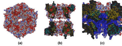
[[Image:Figure_S6.png|left|400px|thumb|Electrostatic potential mapped at the molecular surface for the R1deltaDII/R2deltaDII dodecamer. (a) top view and (b) cross-section view showing the central channel. (c) cross-section view of the SV40 Ltag | [[Image:Figure_S6.png|left|400px|thumb|Electrostatic potential mapped at the molecular surface for the R1deltaDII/R2deltaDII dodecamer. (a) top view and (b) cross-section view showing the central channel. (c) cross-section view of the SV40 Ltag | ||
hexamer complexed with ATP (PDB [[1svm]]), included for comparison. In (a) and (b) the R1deltaDII and R2deltaDII monomers are colored gold and cyan, respectively. In (c) the SV40 monomers are alternately colored gold and cyan.]] | hexamer complexed with ATP (PDB [[1svm]]), included for comparison. In (a) and (b) the R1deltaDII and R2deltaDII monomers are colored gold and cyan, respectively. In (c) the SV40 monomers are alternately colored gold and cyan.]] | ||
| - | + | {{Clear}} | |
Our data give new insights into the molecular arrangement of RuvBL1 and RuvBL2 and strongly suggest that in vivo activities of these highly interesting therapeutic drug targets are regulated by cofactors inducing conformational changes via domain II in order to modulate the enzyme complex into its active state. | Our data give new insights into the molecular arrangement of RuvBL1 and RuvBL2 and strongly suggest that in vivo activities of these highly interesting therapeutic drug targets are regulated by cofactors inducing conformational changes via domain II in order to modulate the enzyme complex into its active state. | ||
Revision as of 06:04, 23 August 2011
| |||||||||||
- ↑ Unknown PubmedID noref
- ↑ Matias PM, Gorynia S, Donner P, Carrondo MA. Crystal structure of the human AAA+ protein RuvBL1. J Biol Chem. 2006 Dec 15;281(50):38918-29. Epub 2006 Oct 23. PMID:17060327 doi:10.1074/jbc.M605625200
This page complements a publication in scientific journals and is one of the Proteopedia's Interactive 3D Complement pages. For aditional details please see I3DC.

