Adrenergic receptor
From Proteopedia
| Line 8: | Line 8: | ||
== 3D Structures of Adrenergic receptor == | == 3D Structures of Adrenergic receptor == | ||
| - | ''Update | + | ''Update November 2011'' |
===α-2 adrenergic receptor=== | ===α-2 adrenergic receptor=== | ||
| Line 21: | Line 21: | ||
[[1dep]] – tB1AR peptide - NMR<br /> | [[1dep]] – tB1AR peptide - NMR<br /> | ||
[[2y00]], [[2y02]], [[2y03]], [[2y04]], [[2vt4]] - tB1AR fragment (mutant) + agonist<br /> | [[2y00]], [[2y02]], [[2y03]], [[2y04]], [[2vt4]] - tB1AR fragment (mutant) + agonist<br /> | ||
| - | [[2ycw]], [[2ycx]], [[2ycz]] - tB1AR fragment (mutant) + antagonist | + | [[2ycw]], [[2ycx]], [[2ycz]], [[2ycy]] - tB1AR fragment (mutant) + antagonist |
===β-2 adrenergic receptor=== | ===β-2 adrenergic receptor=== | ||
| Line 30: | Line 30: | ||
[[3d4s]] - hB2AR/T4 lysozyme (mutant) + cholesterol<br /> | [[3d4s]] - hB2AR/T4 lysozyme (mutant) + cholesterol<br /> | ||
[[3ny8]], [[3ny9]], [[3nya]] – hB2AR + agonist<br /> | [[3ny8]], [[3ny9]], [[3nya]] – hB2AR + agonist<br /> | ||
| - | + | [[3sn6]] - hB2AR + cameloid antibody fragment + guanine nucleotide-binding protein G | |
| - | + | ||
[[Category:Topic Page]] | [[Category:Topic Page]] | ||
Revision as of 10:48, 6 November 2011

| |||||||||
| β-2 Adrenergic Receptor, 2rh1 | |||||||||
|---|---|---|---|---|---|---|---|---|---|
| Ligands: | , , , , , , , | ||||||||
| Gene: | ADRB2, ADRB2R, B2AR / E (Enterobacteria phage T4) | ||||||||
| |||||||||
| |||||||||
| Resources: | FirstGlance, OCA, RCSB, PDBsum | ||||||||
| Coordinates: | save as pdb, mmCIF, xml | ||||||||
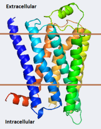
The adrenergic receptors are metabolic G protein-coupled receptors. They are the targets of catecholamines. The binding of an agonist to them causes a sympathetic response. The α-2 adrenergic receptor (A2AR) inhibits insulin or glucagons release. The β-1 adrenergic receptor (B1AR) increases cardiac output and secretion of rennin and ghrelin. The β-2 adrenergic receptor (B2AR) triggers many relaxation reactions. The images at the left and at the right correspond to one representative Adrenergic receptor, i.e. the crystal structure of human β-2 adrenergic Receptor (2rh1). See also Beta-2 Adrenergic Receptor.
Contents |
3D Structures of Adrenergic receptor
Update November 2011
α-2 adrenergic receptor
3kj6, 2r4r, 2r4s – hA2AR + FAB heavy+light chains
1ho9, 1hod – hA2AR peptide (mutant) – NMR
1hof, 1hll - hA2AR peptide – NMR
β-1 adrenergic receptor
2y01 – tB1AR fragment (mutant) – turkey
1dep – tB1AR peptide - NMR
2y00, 2y02, 2y03, 2y04, 2vt4 - tB1AR fragment (mutant) + agonist
2ycw, 2ycx, 2ycz, 2ycy - tB1AR fragment (mutant) + antagonist
β-2 adrenergic receptor
See also Beta-2 Adrenergic Receptor
3pds - hB2AR/T4 lysozyme - human
3p0g – hB2AR/T4 lysozyme + cameloid antibody fragment
2rh1 - hB2AR/T4 lysozyme (mutant)
3d4s - hB2AR/T4 lysozyme (mutant) + cholesterol
3ny8, 3ny9, 3nya – hB2AR + agonist
3sn6 - hB2AR + cameloid antibody fragment + guanine nucleotide-binding protein G
Proteopedia Page Contributors and Editors (what is this?)
Michal Harel, Wayne Decatur, Karsten Theis, Alexander Berchansky

