Lipase, as its name suggests, is an enzyme responsible for the cleavage of types of lipid molecules. There are different types of lipases, many of which work in similar ways. For instance, Human Pancreatic Lipase, or HPL, splits triglycerides, the main lipids in the human diet, into glycerol and three fatty acids. The structure shown at right is that of Horse Pancreatic Lipase. It consists of two identical subunits, totaling 449 amino acids each, and totals 50 kDA. To better visualize the directionality of the subunits with respect to each other we can use a . This diagram shows the N-terminus of each subunit in blue, the follows the spectrum through green, yellow, orange, and finally the C-terminus is shown in red.
Basic Structure
The of lipase (just one subunit) include 102 residues which create 13 alpha helices, shown in red, and 139 residues involved in beta sheets totaling 28 strands, shown in gold. Lipase of course consists of both
. The polar residues in this scene are shown in a light blue shade, and the nonpolar are in a dark red. From this representation, it can be assumed that there is a similar quantity of polar residues as there are nonopolar. The tertiary stucture of the molecule is stabilized by 6 and ionic interactions with a calcium ligand within each subunit. Finally, the quaternary structure is completed by the adjoining of the two identical subunits.The include hydrogen bonds, hydrophobic interactions, salt bridges, and other interactions.
The Calcium Ligand
The are two calcium ions, one buried within each subunit. This scene shows the interactions between the calcium ion (shown in green) in subunit A and the following residues from subunit A: GLU187, ARG190, ASP192, and ASP195. In addition to interactions with these molecules, the calcium ion is also stabilized by the oxygens from two water molecules shown in pink. These interactions between the amino acid residues and the ligand are crucial for proper protein folding, and subsequently protein function.
The Mechanism
Image:Hydrolysis of trigly.png This is the reaction catalyzed by most lipases. Shown is a triglyceride being hydrolyzed by water, resulting in glycerol and three liberated fatty acids. Pancreatic Lipase, however, only hydrolyzes two ester bonds from triglycerides.
Lipase works with a substrate that consists usually of triglycerides. Triglycerides are composed of glycerol connected via ester bonds to three fatty acids. These molecules are split by hydrolysis (see figure). This is the reaction catalyzed by most lipases. Lipase acts on the exterior fatty acids of triglycerides, hydrolyzing the bonds and freeing the two outer fatty acids from the glycerol backbone. The of lipase consists mainly of three residues. These three residues are SER152, ASP176, and HIS263. The mechanism for these hydrolysis reactions begins with SER152 attacking a carbonyl carbon, forming a tetrahedral intermediate. When the oxyanion reforms its double bonded form, the oxygen originally from the ester bond acts as the leaving group and accepts a hydrogen from HIS263. Next, water is converted into a nucleophile by donating a hydrogen to the HIS263, then attacking the new carbonyl. The SER152 now acts as the leaving group, producing a fatty acid chain which has been separated from glycerol and regenerating the enzyme active site.
Evolutionary Conservation
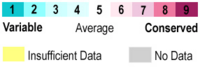
This scale is used to identify the evolutionary conservation of certain residues. The red residues are the most conserved and the blue tend to be variable between variations of the protein.
The consistency of residues between variations of lipases can be described as the of the protein. As it can be observed from the conservation of individual residues, the residues near the active site and close to the calcium ligand have the highest average conservation. The conservation of residues becomes more variable the farther away from each active site.

