This old version of Proteopedia is provided for student assignments while the new version is undergoing repairs. Content and edits done in this old version of Proteopedia after March 1, 2026 will eventually be lost when it is retired in about June of 2026.
Apply for new accounts at the new Proteopedia. Your logins will work in both the old and new versions.
User:Brian Hernandez/DOPA Decarboxylase
From Proteopedia
(Difference between revisions)
(→'''Inhibitor Binding''') |
|||
| Line 7: | Line 7: | ||
==Structure== | ==Structure== | ||
---- | ---- | ||
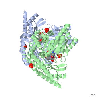
| - | DDC consists of two monomers | + | DDC consists of two monomers, each with three distinct domains: the large domain, the C-terminal small domain, and the N-terminal domain <ref name=Burkhard>PMID: 11685243 </ref>. The <scene name='DOPA_decarboxylase/Large_domain/1'>large domain</scene> consists of the cofactor (PLP- pyridoxal phosphate) binding site and is comprised of a central, seven-stranded mixed β-sheet surrounded by eight α-helices in a typical α/β fold. The small <scene name='DOPA_decarboxylase/Small_domain/1'>C-terminal domain</scene> consists of a four-stranded antiparallel β-sheet with three helices packed against the face opposite the large domain. The <scene name='Sandbox/N-terminal_domain/2'>N-terminal domain</scene> (characteristic of all α-family enzymes) is composed of two parallel helices linked by an extended strand. This structure flaps over the top of the second subunit and vice versa, with the first helix of one subunit aligning parallel to the equivalent helix of the other subunit, thereby forming the extended dimer interface. Although it is unlikely that this domain represents an autonomous folding unit, it is most likely stable only in the context of the dimer by extending the interface between the two monomers. |
==Function== | ==Function== | ||
==='''The Active Site'''=== | ==='''The Active Site'''=== | ||
| - | DDC's active site is located in a <scene name='DOPA_decarboxylase/Dimer_interface/2'>cleft</scene> between the two monomer subunits, but is composed mainly of residues from one monomer.The <scene name='DOPA_decarboxylase/Active_site/1'>active site</scene> is composed of several key residues, including Lys-303, Asp-271, His-192, Thr-82, Ile-101, and Phe-103. In the ligand free form, PLP binds to Lys 303 via a Schiff base linkage. A [http://en.wikipedia.org/wiki/Salt_bridge_(protein) salt bridge] forms between the carboxylate group of Asp 271 and the protonated pyridine nitrogen of PLP yielding a strong electron sink capable of stabilizing the carbanionic intermediates. The only two active site residues from the adjacent monomer, Ile-101 and Phe-103, are part of the substrate binding pocket. | + | DDC's active site is located in a <scene name='DOPA_decarboxylase/Dimer_interface/2'>cleft</scene> between the two monomer subunits, but is composed mainly of residues from one monomer.The <scene name='DOPA_decarboxylase/Active_site/1'>active site</scene> is composed of several key residues, including Lys-303, Asp-271, His-192, Thr-82, Ile-101, and Phe-103. In the ligand free form, PLP binds to Lys 303 via a Schiff base linkage. A [http://en.wikipedia.org/wiki/Salt_bridge_(protein) salt bridge] forms between the carboxylate group of Asp 271 and the protonated pyridine nitrogen of PLP yielding a strong '''electron sink''' capable of stabilizing the carbanionic intermediates. The only two active site residues from the adjacent monomer, Ile-101 and Phe-103, are part of the substrate binding pocket. |
[[image:plp bound.png|thumb|center|400px|'''Schiff base linkage of PLP to Lys303 in the active site''']] | [[image:plp bound.png|thumb|center|400px|'''Schiff base linkage of PLP to Lys303 in the active site''']] | ||
==='''Flexible Loop'''=== | ==='''Flexible Loop'''=== | ||
| - | Residues 328-338 are a stretch of 11 amino acids that comprise a mobile loop (located near the dimer interface), thought to be significant in the catalytic mechanism. During catalysis, the loop is expected to adhere to a less solvent- and protease-exposed conformation, whereby it loses flexibility and extends toward the active site. Tyr-332 and Lys-334 are highly conserved residues indicative of the flexible loop's possible role in catalysis. The conformational change that occurs is thought to be a result of loop residues directly interacting with the inhibitor. Finally, with such a conformational change, the Tyr-332 residue could then be located closer to the substrate to possibly act as a proton donor for the quinonoid Cα. | + | Residues 328-338 are a stretch of 11 amino acids that comprise a mobile loop (located near the dimer interface), thought to be significant in the catalytic mechanism<ref name=Ishii>PMID: 8889823 </ref>. During catalysis, the loop is expected to adhere to a less solvent- and protease-exposed conformation, whereby it loses flexibility and extends toward the active site. Tyr-332 and Lys-334 are highly conserved residues indicative of the flexible loop's possible role in catalysis. The conformational change that occurs is thought to be a result of loop residues directly interacting with the inhibitor. Finally, with such a conformational change, the Tyr-332 residue could then be located closer to the substrate to possibly act as a proton donor for the quinonoid Cα. |
| - | + | ||
| - | + | ||
==DDC and Parkinson's Disease== | ==DDC and Parkinson's Disease== | ||
| Line 26: | Line 24: | ||
The inhibitor <scene name='DOPA_decarboxylase/Carbidopa/1'>carbiDOPA</scene> binds to the enzyme by forming a hydrazone linkage with the PLP cofactor through its hydrazine moiety <ref name=Burkhard>PMID: 11685243 </ref>. | The inhibitor <scene name='DOPA_decarboxylase/Carbidopa/1'>carbiDOPA</scene> binds to the enzyme by forming a hydrazone linkage with the PLP cofactor through its hydrazine moiety <ref name=Burkhard>PMID: 11685243 </ref>. | ||
[[Image:carbidopa.jpeg|thumb|right|400px|'''carbiDOPA''']] The catechol ring buries itself deep within the active site cleft, with the 4' hydroxyl of the catechol ring hydrogen bonded to the hydroxyl group of Thr 82. Additionally, the highly conserved His 192 residue forms a hydrogen bond to the carboxylate group of carbiDOPA (with the carboxylate group of L-DOPA being even closer in binding range). Thus, His 192 is most likely a key residue in the enzyme's catalytic activity because a H192A mutation can cause complete loss of activity. | [[Image:carbidopa.jpeg|thumb|right|400px|'''carbiDOPA''']] The catechol ring buries itself deep within the active site cleft, with the 4' hydroxyl of the catechol ring hydrogen bonded to the hydroxyl group of Thr 82. Additionally, the highly conserved His 192 residue forms a hydrogen bond to the carboxylate group of carbiDOPA (with the carboxylate group of L-DOPA being even closer in binding range). Thus, His 192 is most likely a key residue in the enzyme's catalytic activity because a H192A mutation can cause complete loss of activity. | ||
| + | </StructureSection> | ||
| + | |||
| + | ==3D structures of DOPA decarboxylase== | ||
| + | |||
| + | |||
| + | |||
| + | [[3k40]] – DDC – ''Drosophila melanogaster''<br /> | ||
| + | [[1js3]] – pDDC + inhibitor – pig<br /> | ||
| + | [[1js6]] - pDDC<br /> | ||
| + | [[3rbf]], [[3rbl]] – hDDC – human<br /> | ||
| + | [[3rch]] – hDDC + vitamin B6 phosphate + pyridoxal phosphate | ||
Revision as of 06:23, 29 November 2011
| |||||||||||


3D structures of DOPA decarboxylase
3k40 – DDC – Drosophila melanogaster
1js3 – pDDC + inhibitor – pig
1js6 - pDDC
3rbf, 3rbl – hDDC – human
3rch – hDDC + vitamin B6 phosphate + pyridoxal phosphate
