Image:CTSBFig1.jpg
From Proteopedia
(Difference between revisions)
No higher resolution available.
CTSBFig1.jpg (371 × 350 pixel, file size: 148 KB, MIME type: image/jpeg)
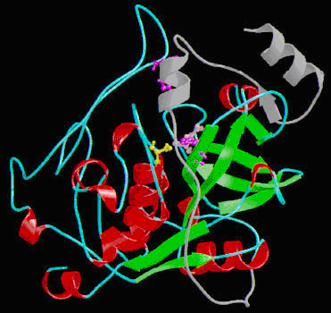
(A diagram representation of Cathepsin B structure: Alpha-helixes are shown in red and β-sheets in green. Catalytic residues are shown in ball-and-stick representation: Cys108 in yellow, His278 in purple and Asn298 in pink. His189 on the occluding loop is) |
(uploaded a new version of "Image:CTSBFig1.jpg": Cathepsin B) |
Current revision
A diagram representation of Cathepsin B structure: Alpha-helixes are shown in red and β-sheets in green. Catalytic residues are shown in ball-and-stick representation: Cys108 in yellow, His278 in purple and Asn298 in pink. His189 on the occluding loop is shown in purple in ball-and-stick representation. The pro-peptide is shown in grey.
File history
Click on a date/time to view the file as it appeared at that time.
| Date/Time | User | Dimensions | File size | Comment | |
|---|---|---|---|---|---|
| (current) | 23:04, 2 May 2012 | Alia Raja (Talk | contribs) | 371×350 | 148 KB | Cathepsin B |
| 18:29, 1 May 2012 | Alia Raja (Talk | contribs) | 371×350 | 148 KB | A diagram representation of Cathepsin B structure: Alpha-helixes are shown in red and β-sheets in green. Catalytic residues are shown in ball-and-stick representation: Cys108 in yellow, His278 in purple and Asn298 in pink. His189 on the occluding loop is |
- Edit this file using an external application
See the setup instructions for more information.
Links
The following pages link to this file:
Metadata
This file contains additional information, probably added from the digital camera or scanner used to create or digitize it. If the file has been modified from its original state, some details may not fully reflect the modified image.
| Orientation | Normal |
|---|---|
| Horizontal resolution | 72 dpi |
| Vertical resolution | 72 dpi |
| Software used | Adobe Photoshop CS2 Windows |
| File change date and time | 16:51, 6 May 2008 |
| Color space | sRGB |
