Image:Neuron Specific Enolase.jpg
From Proteopedia
No higher resolution available.
Neuron_Specific_Enolase.jpg (500 × 500 pixel, file size: 61 KB, MIME type: image/jpeg)
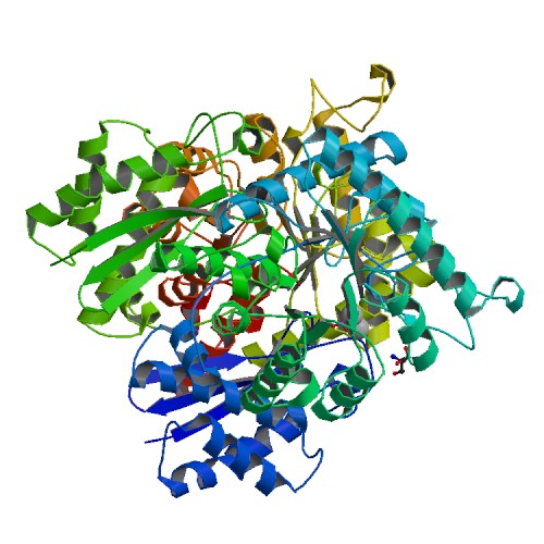
(Biological Assembly Image for 1TE6 Crystal Structure of Human Neuron Specific Enolase at 1.8 angstrom) |
|||
| Line 1: | Line 1: | ||
== Summary == | == Summary == | ||
| - | + | PubMed Abstract: | |
| - | + | Human neuron-specific enolase (NSE) or isozyme gamma has been expressed with a C-terminal His-tag in Escherichia coli. The enzyme has been purified, crystallized and its crystal structure determined. In the crystals the enzyme forms the asymmetric complex NSE x Mg2 x SO4/NSE x Mg x Cl, where "/" separates the dimer subunits. The subunit that contains the sulfate (or phosphate) ion and two magnesium ions is in the closed conformation observed in enolase complexes with the substrate or its analogues; the other subunit is in the open conformation observed in enolase subunits without bound substrate or analogues. This indicates negative cooperativity for ligand binding between subunits. Electrostatic charge differences between isozymes alpha and gamma, -19 at physiological pH, are concentrated in the regions of the molecular surface that are negatively charged in alpha, i.e. surface areas negatively charged in alpha are more negatively charged in gamma, while areas that are neutral or positively charged tend to be charge-conserved. | |
== Licensing == | == Licensing == | ||
{{subst:No license from license selector|Somewebsite}} | {{subst:No license from license selector|Somewebsite}} | ||
Current revision
Summary
PubMed Abstract: Human neuron-specific enolase (NSE) or isozyme gamma has been expressed with a C-terminal His-tag in Escherichia coli. The enzyme has been purified, crystallized and its crystal structure determined. In the crystals the enzyme forms the asymmetric complex NSE x Mg2 x SO4/NSE x Mg x Cl, where "/" separates the dimer subunits. The subunit that contains the sulfate (or phosphate) ion and two magnesium ions is in the closed conformation observed in enolase complexes with the substrate or its analogues; the other subunit is in the open conformation observed in enolase subunits without bound substrate or analogues. This indicates negative cooperativity for ligand binding between subunits. Electrostatic charge differences between isozymes alpha and gamma, -19 at physiological pH, are concentrated in the regions of the molecular surface that are negatively charged in alpha, i.e. surface areas negatively charged in alpha are more negatively charged in gamma, while areas that are neutral or positively charged tend to be charge-conserved.
Licensing
{{subst:No license from license selector|Somewebsite}}
File history
Click on a date/time to view the file as it appeared at that time.
| Date/Time | User | Dimensions | File size | Comment | |
|---|---|---|---|---|---|
| (current) | 04:58, 8 May 2012 | William J. Barnes (Talk | contribs) | 500×500 | 61 KB | Biological Assembly Image for 1TE6 Crystal Structure of Human Neuron Specific Enolase at 1.8 angstrom |
- Edit this file using an external application
See the setup instructions for more information.
Links
The following pages link to this file:
Metadata
This file contains additional information, probably added from the digital camera or scanner used to create or digitize it. If the file has been modified from its original state, some details may not fully reflect the modified image.
| Image title | ImageSource=RCSB PDB; StructureID=1te6; DOI=http://dx.doi.org/10.2210/pdb1te6/pdb; |
|---|
