We apologize for Proteopedia being slow to respond. For the past two years, a new implementation of Proteopedia has been being built. Soon, it will replace this 18-year old system. All existing content will be moved to the new system at a date that will be announced here.
2mad
From Proteopedia
(Difference between revisions)
(New page: 200px <!-- The line below this paragraph, containing "STRUCTURE_2mad", creates the "Structure Box" on the page. You may change the PDB parameter (which sets the PD...) |
m (Protected "2mad" [edit=sysop:move=sysop]) |
Revision as of 10:02, 2 June 2012
| |||||||||
| 2mad, resolution 2.25Å () | |||||||||
|---|---|---|---|---|---|---|---|---|---|
| Non-Standard Residues: | |||||||||
| Activity: | Amine dehydrogenase, with EC number 1.4.99.3 | ||||||||
| |||||||||
| |||||||||
| Resources: | FirstGlance, OCA, PDBsum, RCSB | ||||||||
| Coordinates: | save as pdb, mmCIF, xml | ||||||||
Contents |
THE ACTIVE SITE STRUCTURE OF METHYLAMINE DEHYDROGENASE: HYDRAZINES IDENTIFY C6 AS THE REACTIVE SITE OF THE TRYPTOPHAN DERIVED QUINONE COFACTOR
Template:ABSTRACT PUBMED 1390754
About this Structure
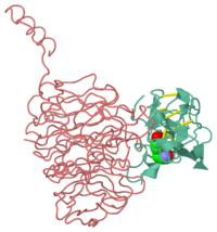
2mad is a 2 chain structure of Methylamine dehydrogenase with sequence from Paracoccus versutus. This structure supersedes the now removed PDB entry 1mad. Full crystallographic information is available from OCA.
See Also
Reference
- Huizinga EG, van Zanten BA, Duine JA, Jongejan JA, Huitema F, Wilson KS, Hol WG. Active site structure of methylamine dehydrogenase: hydrazines identify C6 as the reactive site of the tryptophan-derived quinone cofactor. Biochemistry. 1992 Oct 13;31(40):9789-95. PMID:1390754


