Transcription-repair coupling factor
From Proteopedia
| Line 1: | Line 1: | ||
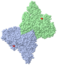
[[Image:2eyq.png|left|200px|thumb|Crystal Structure of Transcription-repair coupling factor [[2eyq]]]] | [[Image:2eyq.png|left|200px|thumb|Crystal Structure of Transcription-repair coupling factor [[2eyq]]]] | ||
{{STRUCTURE_2eyq| PDB=2eyq | SCENE=2eyq/Asymmunit/2 |CAPTION= 2eyq asymmetric unit, resolution 3.20Å. <br> Click <scene name='2eyq/Biolunit/1'>here</scene> to display biological molecule.}} | {{STRUCTURE_2eyq| PDB=2eyq | SCENE=2eyq/Asymmunit/2 |CAPTION= 2eyq asymmetric unit, resolution 3.20Å. <br> Click <scene name='2eyq/Biolunit/1'>here</scene> to display biological molecule.}} | ||
| - | 2EYQ is a 2 chains structure of sequences from [http://en.wikipedia.org/wiki/Escherichia_coli Escherichia coli]. Full crystallographic information is available from [http://oca.weizmann.ac.il/oca-bin/ocashort?id=2EYQ OCA]. | ||
| - | |||
'''Transcription-repair coupling factor (TRCF)''' enables the coupling of these processes in bacteria and humans. The TRCF has ATPase activity. <scene name='2eyq/Asymmunit/2'>The crystallographic asymmetric unit of the solved structure contains 2 monomers of Mfd (residues 2-1147 of 1151 for one molecule and 5-1147 of the other), five HEPES molecules, and the sulfate ions.</scene> | '''Transcription-repair coupling factor (TRCF)''' enables the coupling of these processes in bacteria and humans. The TRCF has ATPase activity. <scene name='2eyq/Asymmunit/2'>The crystallographic asymmetric unit of the solved structure contains 2 monomers of Mfd (residues 2-1147 of 1151 for one molecule and 5-1147 of the other), five HEPES molecules, and the sulfate ions.</scene> | ||
Revision as of 12:56, 6 June 2012

Template:STRUCTURE 2eyq Transcription-repair coupling factor (TRCF) enables the coupling of these processes in bacteria and humans. The TRCF has ATPase activity.
Size exclusion chromatogaphy indicates that the protein
an N→C rainbow similar to Figure 1A of the article describing the structure. .
The domains in the structure of Mfd:
similar to Figure 1B and Figure 2 of the article describing the structure.
Figure 1B and Figure 2 of the article describing the structure. .
[Note: the following view generates a substantial surface which may take several minutes to calculate. Use the one above as an alternative unless you are willing to spend the time.] Figure 1B. .
displayed as in Figure 3A of the article describing the structure.
are highlighted in red. See figure 3B.
displayed similar to Figure 3B of the article describing the structure.
indicated similar to Figure 3C of the article describing the structure.
3D Structures of Transcription-repair coupling factor
Updated December 2011
2eyq, 2b2n – EcTRCF – Escherichia coli
3hjh – EcTRCF residues 1-470
3mlq – TRCF RNA polymerase interacting domain + DNA-directed RNA polymerase subunit β - Thermus thermophilus
2qsr – TRCF C terminal – Streptococcus pneumoni
Reference
- Deaconescu AM, Chambers AL, Smith AJ, Nickels BE, Hochschild A, Savery NJ, Darst SA. Structural basis for bacterial transcription-coupled DNA repair. Cell. 2006 Feb 10;124(3):507-20. PMID:16469698 doi:10.1016/j.cell.2005.11.045
Created with the participation of Wayne Decatur
Proteopedia Page Contributors and Editors (what is this?)
Karsten Theis, Michal Harel, Alexander Berchansky, Wayne Decatur
