Sandbox christian
From Proteopedia
| Line 12: | Line 12: | ||
<StructureSection load='1tca' size='600' side='right' caption='Structure of CALB (PDB entry [[1tca]])' scene=''> | <StructureSection load='1tca' size='600' side='right' caption='Structure of CALB (PDB entry [[1tca]])' scene=''> | ||
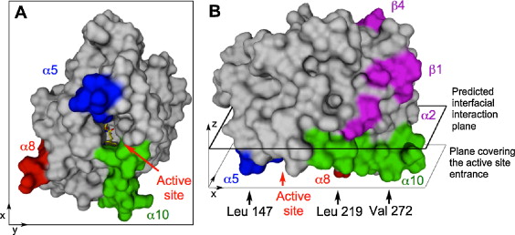
| - | The Active Site consists of <scene name='Sandbox_christian/Active_site/1'> | + | The Active Site consists of <scene name='Sandbox_christian/Active_site/1'>Ser 105, Asp 187 and His 224</scene>. Click here to see the <scene name='Sandbox_christian/Active_site/2'> B-factors</scene> and the <scene name='Sandbox_christian/Active_site/3'>Glycosylation site</scene>. The above described hydrophobic anchors are <scene name='Sandbox_christian/Active_site/5'>Leu 147, Leu 219, and Val 272</scene>. |
</StructureSection> | </StructureSection> | ||
<References/> | <References/> | ||
Revision as of 11:27, 6 September 2012
Candida antarctica lipase B
Lipase B from Candida antarctica binds to hydrophobic substrate–water interfaces via hydrophobic anchors surrounding the active site entrance [1]
Abstract: Candida antarctica Lipase B (CALB) has been extensively studied over the past decades and has proved to be an efficient catalyst for various industrial and scientific applications. Because of its ability to hydrolyse soluble and insoluble substrates and the lack of a classical interfacial activation it was previously characterized as an intermediate between a lipase and an esterase. We show by molecular dynamics simulation in full atomistic detail that CALB attaches and binds to a hydrophobic tributyrin–water interface via three hydrophobic anchor regions defined by Leu 147, Leu 219, and Val 272 surrounding the entrance to the active site. These regions trigger the reorientation of the protein via hydrophobic interactions even when the protein impacts at the surface in a non-optimal orientation. During the binding process the flexible helix α5 undergoes a movement of 7.5 Å towards the substrate layer. Though tributyrin has no net charge a long-range attraction between interface and protein was observed up to a separation of 7 Å that corresponds to the amplitude of the fluctuations of the tributyrin surface. A stable binding of the protein with the active site oriented towards the active site was observed for several hundreds of nanoseconds in total. During that time, single tributyrin molecules moved from the tributyrin layer into the substrate binding site of CALB and were temporarily binding as productive sn3-complexes.

(A) Hydrophobic regions of CALB: anchor residues Leu 147, Leu 219, and Val 272 are located in helix α5 (blue), α 8 (red) and α 10 (green) surrounding the entrance to the active site (Ser 105, His 224 and Trp 104 shown as yellow sticks). (B) The outermost hydrophobic residues Leu 147, Leu 219, and Val 272 define the x/y-plane covering the active site (thin line). The interfacial interaction plane predicted by the “Orientations of Proteins in Membranes” (OPM) database is parallel to this x/y plane (thick line).
| |||||||||||
- ↑ C. Gruber, J. Pleiss: Lipase B from Candida antarctica binds to hydrophobic substrate-water interfaces via hydrophobic anchors surrounding the active site entrance, J MOL CATAL B-ENZYM, 2012; http://dx.doi.org/10.1016/j.molcatb.2012.05.012
