Group:MUZIC:Telethonin
From Proteopedia
(Difference between revisions)

(→Telethonin) |
|||
| Line 1: | Line 1: | ||
= Telethonin = | = Telethonin = | ||
<StructureSection load='1ya5' size='300' side='left' caption='Telethonin crystal structure by Zou et al. (2006) interacting with Z1 and Z2 titin domains(PDB entry [[1ya5]])' scene='User:Marcia_Ivonne_Pena_Paz/workbench/Telethonin/Telethonin1/2'> | <StructureSection load='1ya5' size='300' side='left' caption='Telethonin crystal structure by Zou et al. (2006) interacting with Z1 and Z2 titin domains(PDB entry [[1ya5]])' scene='User:Marcia_Ivonne_Pena_Paz/workbench/Telethonin/Telethonin1/2'> | ||
| - | Also known as T-Cap or Titin Cap protein. | + | Also known as T-Cap or Titin Cap protein. |
| - | + | ||
| - | + | ||
| - | + | ||
| - | + | ||
| - | + | ||
| - | + | ||
| + | == Introduction == | ||
| + | It is a small protein of 19kDa, 167 amino acids. It is predominantly expressed in striated muscle. It is a structural protein of the muscle; it is associated to the Z-disc in the sarcomere. It acts as multifunctional protein linking titin and other proteins implicated in sarcomere structure and signalling pathways. | ||
It is encoded by the ''Tcap'' gene, in mice (''Mus musculus'') and ''TCAP'' in humans (''Homo sapiens''). | It is encoded by the ''Tcap'' gene, in mice (''Mus musculus'') and ''TCAP'' in humans (''Homo sapiens''). | ||
| Line 21: | Line 17: | ||
| + | == Sequence Annotation == | ||
| + | Telethonin is comprised of 167 amino acids, the sequence of [http://http://www.uniprot.org/uniprot/O15273 human], [http://www.uniprot.org/uniprot/O70548 mouse], [http://www.uniprot.org/uniprot/Q6T8D8 bovine], [http://www.uniprot.org/uniprot/A4GR69 porcine] telethonin is available from Uniprot. | ||
| - | == ''Tcap'' gene product, the protein Telethonin == | ||
| + | |||
| + | |||
| + | == Structure == | ||
Telethonin protein is found mostly in skeletal and cardiac muscle. It is one of the major components of the sarcomere, it is localized to the Z-disc. It was also reported a nuclear localization.<ref>PMID:12379311 </ref>, <ref name="a"> PMID:16678796 </ref> | Telethonin protein is found mostly in skeletal and cardiac muscle. It is one of the major components of the sarcomere, it is localized to the Z-disc. It was also reported a nuclear localization.<ref>PMID:12379311 </ref>, <ref name="a"> PMID:16678796 </ref> | ||
| - | Studies on telethonin structure by Zou et al. <ref name="b"> PMID:16407954 </ref> report that it is made up of <scene name='User:Marcia_Ivonne_Pena_Paz/workbench/Telethonin/Telethonin_nter_cter/1'>five stranded antiparallel β-sheets extended by two wing-shaped β-hairpin motifs (A-B, C-D). These two motifs are related by an approximate two-fold symmetry, which generates an almost perfect palindromic arrangement.</scene> (N-terminal in blue and C-ter in orange) | + | Studies on telethonin structure by Zou et al. <ref name="b"> PMID:16407954 </ref> report that it is made up of <scene name='User:Marcia_Ivonne_Pena_Paz/workbench/Telethonin/Telethonin_nter_cter/1'>five stranded antiparallel β-sheets extended by two wing-shaped β-hairpin motifs (A-B, C-D). These two motifs are related by an approximate two-fold symmetry, which generates an almost perfect palindromic arrangement.</scene> (N-terminal in blue and C-ter in orange). |
The structure of telethonin was determined using X-ray crystallography. <ref>PMID:12446666</ref>,<ref name="b" /> The shape and architecture of the complex of titin/telethonin was studied by small-angle- X-ray scattering (SAXS) and then compared to the crystallographic models. They also used in-vitro experiments to follow the formation of the complex in non-myogenic Cos1 cells, in order to understand if the assemblage is possible <ref>PMID:16713295</ref> | The structure of telethonin was determined using X-ray crystallography. <ref>PMID:12446666</ref>,<ref name="b" /> The shape and architecture of the complex of titin/telethonin was studied by small-angle- X-ray scattering (SAXS) and then compared to the crystallographic models. They also used in-vitro experiments to follow the formation of the complex in non-myogenic Cos1 cells, in order to understand if the assemblage is possible <ref>PMID:16713295</ref> | ||
| - | This symmetry of telethonin permits its interaction with titin. Both are assembled in an antiparallel <scene name='User:Marcia_Ivonne_Pena_Paz/workbench/Telethonin/Telethonin-titin/1'>sandwich 2:1</scene> (titin:telethonin). Titin N-terminal domains Z1 and Z2 (two Ig like repeats) interact with the C-terminal region of telethonin (residues 1-53). Telethonin mediates in the antiparallel assembly of the two Z1Z2domains. | + | This symmetry of telethonin permits its interaction with titin. Both are assembled in an antiparallel <scene name='User:Marcia_Ivonne_Pena_Paz/workbench/Telethonin/Telethonin-titin/1'>sandwich 2:1</scene> (titin:telethonin). Titin N-terminal domains Z1 and Z2 (two Ig like repeats) interact with the C-terminal region of telethonin (residues 1-53). Telethonin mediates in the antiparallel assembly of the two Z1Z2domains. This structure is only found in the presence of titin, it might adopt a different fold in its absence. |
| - | In early differentiating myocytes titin C-terminal and telethonin co-localize and titin kinase is close to telethonin C-terminal, and it is phosphorylated. This phosphorylation is involved in the reorganization of the cytoskeleton during myofibrillogenesis. <ref name="c"> PMID:9804419 </ref> This co-localization is not seen in adult myofibrils, titin kinase is reported to localize in the M-band <ref name="c" />; It was also informed that telethonin interacts with other proteins including: Potassium channel β-subunit of the slow activating component of the delayed rectifier potassium current (IKs) channel (minK) <ref name="d"> PMID:11697903 </ref>, ankyrin1, and Z-disc proteins FATZ,/Myozenin-1/ Calsarcin-3 <ref name="e"> PMID:11842093 </ref>, and Ankrd2.<ref name="f"> PMID:15136035 </ref> | ||
| - | Telethonin interacts with minK’s cytoplasmic domain. MinK binds specifically to the sixteen C-terminal residues of telethonin. This suggest that minK, telethonin ant titin form a complex that links myofibrils to the sarcolemma. Phosphorilation of telethonin in Ser157 is a negative regulation for this interaction. This interaction occurs in cardiac myofibrils, it has been reported that minK is not expressed in skeletal muscle. <ref name="d" /> | + | |
| + | |||
| + | == Function and Interactions == | ||
| + | |||
| + | |||
| + | In early differentiating myocytes titin C-terminal and telethonin co-localize and titin kinase is close to telethonin C-terminal, and it is phosphorylated. This phosphorylation is involved in the reorganization of the cytoskeleton during myofibrillogenesis. <ref name="c"> PMID:9804419 </ref> This co-localization is not seen in adult myofibrils, titin kinase is reported to localize in the M-band <ref name="c" />; It was also informed that telethonin interacts with other proteins including: Potassium channel β-subunit of the slow activating component of the delayed rectifier potassium current (IKs) channel (minK) <ref name="d"> PMID:11697903 </ref>, ankyrin1 <ref>PMID:12444090 </ref>, and Z-disc proteins FATZ,/Myozenin-1/ Calsarcin-3 <ref name="e"> PMID:11842093 </ref>, and Ankrd2.<ref name="f"> PMID:15136035 </ref> | ||
| + | |||
| + | Telethonin interacts with minK’s cytoplasmic domain. MinK binds specifically to the sixteen C-terminal residues of telethonin. This suggest that minK, telethonin ant titin form a complex that links myofibrils to the sarcolemma. Phosphorilation of telethonin in Ser157 is a negative regulation for this interaction. This interaction occurs in cardiac myofibrils, it has been reported that minK is not expressed in skeletal muscle. <ref name="d" />. | ||
Telethonin interacts with FATZ/Myozenin-1/Calsarcin-3 N-terminal between residues 78-125. It might be an association as mechanosensing and stretch-associated signalling machinery. <ref name="e" /> | Telethonin interacts with FATZ/Myozenin-1/Calsarcin-3 N-terminal between residues 78-125. It might be an association as mechanosensing and stretch-associated signalling machinery. <ref name="e" /> | ||
| Line 43: | Line 49: | ||
The interaction between Ankrd2 and telethonin has been proposed as a sensor of muscle stress/stretch and a starting point for the transmission of the mechanical signal to the nucleus regulating gene expression. <ref name="f" /> | The interaction between Ankrd2 and telethonin has been proposed as a sensor of muscle stress/stretch and a starting point for the transmission of the mechanical signal to the nucleus regulating gene expression. <ref name="f" /> | ||
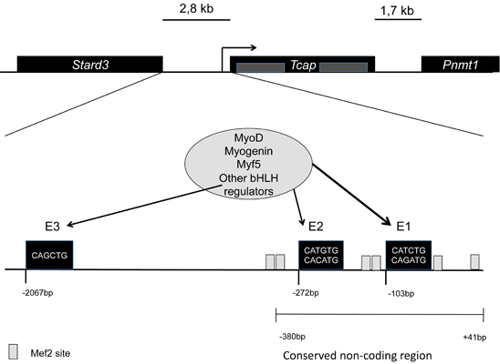
| - | Telethonin is also involved in signalling processes that regulate muscle development. A feed back loop is formed with MRFs (MyoD, myogenin, Myf5) regulating Tcap gene expression; telethonin interacts with myostatin, inhibiting it. So it regulates MyoD through the Myostatin – Smad3 pathway. <ref>PMID:18440815</ref>. | + | Telethonin is also involved in signalling processes that regulate muscle development. A feed back loop is formed with MRFs (MyoD, myogenin, Myf5) regulating Tcap gene expression; telethonin interacts with myostatin, inhibiting it. So it regulates MyoD through the Myostatin – Smad3 pathway. <ref>PMID:18440815</ref>. The interaction with mature Myostatin only occurs with full length Telethonin occuring either in the cytoplasm or the Golgi, it is reported that Telethonin is a negative regulator of myostatin. <ref>PMID:12209887</ref> |
There is an interaction with MDM2 N-terminal. MDM2 is capable of redirecting telethonin to the nucleus. Telethonin is inhibited by MDM2 in a dose dependent manner. In cells MDM2 is involved in the regulation of proteasomal turnover of telethonin. <ref name="a" /> | There is an interaction with MDM2 N-terminal. MDM2 is capable of redirecting telethonin to the nucleus. Telethonin is inhibited by MDM2 in a dose dependent manner. In cells MDM2 is involved in the regulation of proteasomal turnover of telethonin. <ref name="a" /> | ||
| Line 49: | Line 55: | ||
Another interaction has been reported, and also associated with pathology, the one with bone morphogenetic protein-10 (BMP10). The interaction of telethonin with BMP10 is described as a sensor of increased wall stress of the left ventricle. A BMP10 variant is associated with hypertension dilated cardiomyopathy; its binding to telethonin is reduced, and its extracellular secretion is increased, causing cardiomyocyte hypertrophy. <ref>PMID:17921333 </ref> | Another interaction has been reported, and also associated with pathology, the one with bone morphogenetic protein-10 (BMP10). The interaction of telethonin with BMP10 is described as a sensor of increased wall stress of the left ventricle. A BMP10 variant is associated with hypertension dilated cardiomyopathy; its binding to telethonin is reduced, and its extracellular secretion is increased, causing cardiomyocyte hypertrophy. <ref>PMID:17921333 </ref> | ||
| + | Yeast two hybrid screens of skeletal muscle cDNA libraries with baits for the E3 ubiquitin ligases MURF1 and MURF2 have shown a posible targeting of telethonin. <ref>PMID:15967462 </ref> | ||
| + | It was also shown by Y2H an interaction of the proapototic protein Siva and Telethonin, it verified by in vitro pull-down assays, and immunoflurescence experiments showed a colocalization of both proteins in transfected HEK293 cells, but not in vivo. <ref>PMID:18849585</ref> | ||
| - | + | Protein Kinase D (PKD) catalytic domain interacts with Telethonin. It was shown that Telethonin has a PKD recognition motif Arg-X-X-Ser. PKD might regulate sarcomeric assembly and turnover through phosphorylation of Telethonin. <ref>PMID: 155114163 </ref> | |
| - | + | ||
== Pathologies associated with telethonin == | == Pathologies associated with telethonin == | ||
| - | |||
| - | |||
Different mutations in telethonin have been associated with several myopathies. Mutations can lead to limb-girdle muscular dystrophy type 2G (LGMD2G) <ref name="g"> PMID:10655062 </ref>, to hypertrophic cardiopathy, <ref name="h"> PMID:12507422 </ref> and dilated cardiomyopathy. | Different mutations in telethonin have been associated with several myopathies. Mutations can lead to limb-girdle muscular dystrophy type 2G (LGMD2G) <ref name="g"> PMID:10655062 </ref>, to hypertrophic cardiopathy, <ref name="h"> PMID:12507422 </ref> and dilated cardiomyopathy. | ||
| Line 64: | Line 69: | ||
Defects in the MLP-Tcap association are linked to human dilated cardiomyopathy and heart failure (Knöll 2002). Mutations that affect ability of MLP to interact with telethonin result in the loss of telethonin binding, facilitating its mislocalization from the complex with titin, leading to defects in the Z-disc and progression of dilated cardiomyopathy. Knöll et al. conclude that genetic mutations causing a incorrect interaction of telethonin with MLP can lead to a development of human dilated cardiomyopathy through modifications in the conformation and function of titin. <ref name="h" /> | Defects in the MLP-Tcap association are linked to human dilated cardiomyopathy and heart failure (Knöll 2002). Mutations that affect ability of MLP to interact with telethonin result in the loss of telethonin binding, facilitating its mislocalization from the complex with titin, leading to defects in the Z-disc and progression of dilated cardiomyopathy. Knöll et al. conclude that genetic mutations causing a incorrect interaction of telethonin with MLP can lead to a development of human dilated cardiomyopathy through modifications in the conformation and function of titin. <ref name="h" /> | ||
| - | It was reported that in 10 cases of neurogenic atrophy there was a decreased staining for telethonin in type II fibers, and in early stages of fiber atrophy, <ref>PMID: 11763198</ref> indicating a selective downregulation of telethonin. These observations can be related to in vivo studies done in rats, in which after short term denervation (two days), Tcap transcript is reduced by about 50% in skeletal muscle. <ref name="Mason" />. | + | It was reported that in 10 cases of neurogenic atrophy there was a decreased staining for telethonin in type II fibers, and in early stages of fiber atrophy, <ref>PMID: 11763198</ref> indicating a selective downregulation of telethonin. These observations can be related to in vivo studies done in rats, in which after short term denervation (two days), Tcap transcript is reduced by about 50% in skeletal muscle. <ref name="Mason" />. References |
| - | + | ||
| - | + | ||
| - | + | ||
| - | + | ||
| - | + | ||
| - | + | ||
| - | + | ||
| - | + | ||
| - | + | ||
Revision as of 09:39, 25 October 2012
Telethonin
| |||||||||||

