Herceptin - Mechanism of Action
From Proteopedia
| Line 3: | Line 3: | ||
== '''Basics''' == | == '''Basics''' == | ||
=== Introduction === | === Introduction === | ||
| - | Breast cancer is the most common type of cancer found among women. Although it is rarely seen in men, one in eight women will be diagnosed with breast cancer within their lifetime. Patients exhibiting an over-expression in Human Epidermal Growth Factor Receptor 2 (HER2) account for 25% of all breast cancer. HER2+ patients often experience a more aggressive cancer resulting in more metastasized tumors. The statistics show a poor prognosis for HER2+ patients with a 5-year survival rate of 68%. Herceptin (also known as trastuzumab) was approved by the FDA in September of 1998 for HER2+ patients and has been shown to be an effective tool in the battle against breast cancer. | + | Breast cancer is the most common type of cancer found among women except for skin cancers. Although it is rarely seen in men, one in eight women will be diagnosed with breast cancer within their lifetime. Patients exhibiting an over-expression in Human Epidermal Growth Factor Receptor 2 (HER2) account for 25% of all breast cancer. HER2+ patients often experience a more aggressive cancer resulting in more metastasized tumors. The statistics show a poor prognosis for HER2+ patients with a 5-year survival rate of 68%. Herceptin (also known as trastuzumab) was approved by the FDA in September of 1998 for HER2+ patients and has been shown to be an effective tool in the battle against breast cancer. |
=== HER2 === | === HER2 === | ||
| Line 22: | Line 22: | ||
''ERBB2'' is located on the long arm of human chromosome 17 (17q12). This gene codes for the protein HER2 and is known as a proto-oncogene due to its ability to become an oncogene from an over-expression. | ''ERBB2'' is located on the long arm of human chromosome 17 (17q12). This gene codes for the protein HER2 and is known as a proto-oncogene due to its ability to become an oncogene from an over-expression. | ||
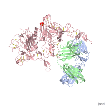
| - | The HER family is composed of three domains: an <scene name='Sandbox_reserved_Herceptin/Extracellular_domain/2'>extracellular domain</scene> further divided into four sub-domains composed of about 630 amino acids, a <scene name='Sandbox_reserved_Herceptin/Transmembrane_domain/1'>transmembrane domain</scene> composed of a single membrane-spanning region, and an <scene name='Sandbox_reserved_Herceptin/Intracellular_domain/1'>intracellular domain</scene> composed of a tyrosine kinase. The four | + | The HER family is composed of three domains: an <scene name='Sandbox_reserved_Herceptin/Extracellular_domain/2'>extracellular domain</scene> further divided into four sub-domains composed of about 630 amino acids, a <scene name='Sandbox_reserved_Herceptin/Transmembrane_domain/1'>transmembrane domain</scene> composed of a single membrane-spanning region, and an <scene name='Sandbox_reserved_Herceptin/Intracellular_domain/1'>intracellular domain</scene> composed of a tyrosine kinase. The four subdomains of the extracellular region include: |
| - | *<scene name='Sandbox_reserved_Herceptin/ | + | *<scene name='Sandbox_reserved_Herceptin/Subdomain_i/2'>sub-domain I</scene> |
| - | *<scene name='Sandbox_reserved_Herceptin/ | + | *<scene name='Sandbox_reserved_Herceptin/Subdomain_ii/3'>sub-domain II</scene> |
| - | *<scene name='Sandbox_reserved_Herceptin/ | + | *<scene name='Sandbox_reserved_Herceptin/Subdomain_iii/3'>sub-domain III</scene> |
| - | *<scene name='Sandbox_reserved_Herceptin/ | + | *<scene name='Sandbox_reserved_Herceptin/Subdomain_iv/3'>sub-domain IV</scene> |
| - | In EGFR, HER3, and HER4 sub-domains I and III remain in a “closed” conformation until a ligand binds. | + | In EGFR, HER3, and HER4 sub-domains I and III remain in a “closed” conformation until a ligand binds. Subdomain II remains hidden making contact with sub-domain IV creating a "tether" (See figure 6). The tether between sub-domain II and IV can be temporarily broken in order for the receptor to become more available for a ligand to bind. Although unsure, evidence suggests that the ligand binds to subdomain I and/or subdomain III; also, there’s some evidence of ligand-binding to subdomain IV. Once ligand-binding is initiated, the receptor experiences a conformational change allowing sub-domains I and III to become closer together resulting in the “open” conformation. This open conformation exposes subdomain II which allows for dimerization between receptors. Although some evidence suggests that sub-domain IV is necessary for ligand-binding, stabilizing the extracellular domain, and locking the receptor in an open conformation, the exact function is still unknown. Once the dimerization between the two receptors takes place, a cross-phosphorylation reaction between the two tyrosine kinases occurs following the activation of certain cell signaling pathways. These pathways include: |
*mitogen-activated protein kinase (MAPK) | *mitogen-activated protein kinase (MAPK) | ||
*phosphoinositide 3-kinase (PI3K/Akt) | *phosphoinositide 3-kinase (PI3K/Akt) | ||
| Line 36: | Line 36: | ||
[[Image:EGFRs.png|thumb|left|250 px|(Figure 5) HER family structures]] | [[Image:EGFRs.png|thumb|left|250 px|(Figure 5) HER family structures]] | ||
| - | HER2 structure differs in that | + | HER2 structure differs in that subdomains I and III are in constant contact resulting in a permanent open conformation. Subdomains I and III in HER2 are stabilized by core hydrophobic residues surrounded by hydrophilic contacts facilitating the stabilization of this interaction. This fixed conformation allows the receptor to act in a ligand-independent manner and permits the access of subdomain II for dimerization. Perhaps this may explain why a high-affinity ligand for HER2 has not yet been recognized. The absence of subdomain II and IV contact can be explained by the mutations of Gly 563 and His 565 found in the other members of the HER family to Pro and Phe respectively. Since subdomain II is always available for dimerization it is free to form heterodimers with any of the other epidermal growth factor receptors. Asp 285 is the only residue in subdomain II not conserved between EGFR and HER2. Leu replaces Asp 285 in HER2 and can explain why HER2 forms homodimers to a lesser extent. Once dimerized, HER2 activates the MAPK pathway, which in turn initiates cell proliferation, migration, differentiation, and angiogenesis. |
HER2 also has another potentially harmful ability. The juxtamembrane region of the extracellular domain is prone to proteolytic cleavage resulting in a protein called p95HER2. <scene name='Sandbox_reserved_Herceptin/Truncated/1'>p95HER2</scene> is the truncated form of HER2 and has been shown to have an increased ability to cross-phosphorylate with the other receptors of the HER family and has increased cellular signaling capabilities. | HER2 also has another potentially harmful ability. The juxtamembrane region of the extracellular domain is prone to proteolytic cleavage resulting in a protein called p95HER2. <scene name='Sandbox_reserved_Herceptin/Truncated/1'>p95HER2</scene> is the truncated form of HER2 and has been shown to have an increased ability to cross-phosphorylate with the other receptors of the HER family and has increased cellular signaling capabilities. | ||
Revision as of 17:05, 13 November 2012

Basics
Introduction
Breast cancer is the most common type of cancer found among women except for skin cancers. Although it is rarely seen in men, one in eight women will be diagnosed with breast cancer within their lifetime. Patients exhibiting an over-expression in Human Epidermal Growth Factor Receptor 2 (HER2) account for 25% of all breast cancer. HER2+ patients often experience a more aggressive cancer resulting in more metastasized tumors. The statistics show a poor prognosis for HER2+ patients with a 5-year survival rate of 68%. Herceptin (also known as trastuzumab) was approved by the FDA in September of 1998 for HER2+ patients and has been shown to be an effective tool in the battle against breast cancer.
HER2
HER2 is one of four human epidermal growth factor receptors (EGFR , HER2, HER3, and HER4). These receptors are part of a family of receptor tyrosine kinases responsible for cell proliferation and differentiation.
These human epidermal growth factor receptors exist on the cell surface and, with the exception of HER2, bind to specific ligands (epidermal growth factors). Over 11 different ligands for the epidermal growth factor receptors have been identified. After binding with these ligands the HER family is able to homodimerize or heterodimerize with one another. This dimerization causes a cross-phosphorylation of the intracellular tyrosine kinases between the two receptors and ultimately activates a cell signaling pathway.
HER2 is the only receptor within this family that is constitutively active being able to dimerize with other HER family members acting in a ligand-independent manner. This continuous activation of the cell signal pathway causes an increase in cell division; thus, potentially causing a tumor.

Herceptin
Herceptin, generic trastuzumab, is a monoclonal antibody. Herceptin is an effective treatment for breast cancer for the reason that it binds to the extracellular domain of HER2 and, by multiple mechanisms of action, can prevent cell proliferation as well as target these HER2+ cells for destruction by the immune system.
| |||||||||||