Caspase-3 Regulatory Mechanisms
From Proteopedia
(→Caspase-3 Loop Bundle and Active Site) |
|||
| Line 28: | Line 28: | ||
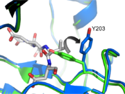
[[Image:Y203flip.png | thumb| Tyrosine 203 Flip]] | [[Image:Y203flip.png | thumb| Tyrosine 203 Flip]] | ||
| - | Taking a closer look at L2 and L2’ we can see a critical interaction involving <scene name='Caspase-3_Regulatory_Mechanisms/D169_nospin/4'>Aspartate 169</scene> on L2. This residue makes two hydrogen bonds with backbone amides of V189’ and E190’, stabilizing L2 in the proper position. This reinforcement allows L2 to contact L3 so as to twist the active site cysteine into the proper orientation to attack the substrate. It also causes a conformational change at Tyrosine 203 (Tyrosine 203 Flip). | + | Taking a closer look at L2 and L2’ we can see a critical interaction involving <scene name='Caspase-3_Regulatory_Mechanisms/D169_nospin/4'>Aspartate 169</scene> on L2. This residue makes two hydrogen bonds with backbone amides of V189’ and E190’ on L2', stabilizing L2 in the proper position. This reinforcement allows L2 to contact L3 so as to twist the active site cysteine into the proper orientation to attack the substrate. It also causes a conformational change at Tyrosine 203 (Tyrosine 203 Flip). Initially, the hydroxyl group on the tyrosine occupies the P1 position in the active site, blocking substrate binding (green, pdb 1QX3). However, when L2 contacts L2' and finds the proper orientation, Y203 rotates 90 degrees (blue, pdb 2H5I) and leaves a hole for P1 of the substrate (substrate shown in gray). In addition, L2 can now contact L4 at K260. This secures L4 and allows it to make contacts in the P4 position, which greatly influence substrate specificity. |
| Line 40: | Line 40: | ||
</StructureSection> | </StructureSection> | ||
| - | |||
== Caspase-3 Regulation== | == Caspase-3 Regulation== | ||
Revision as of 00:42, 13 December 2012
Introduction
Caspases are cysteine-aspartic acid proteases and are key protein facilitators for the faithful execution of apoptosis or programmed cell death. Dysregulation in the apoptotic pathway has been implicated in a variety of diseases such as neurodegeneration, cancer, heart disease and some metabolic disorders. Because of the crucial role of caspases in the the apoptotic pathway, abnormalities in their functions would cause a haywire in the apoptotic cascade and can be deleterious to the cell. Caspases are thus being considered as therapeutic targets in apoptosis-related diseases.
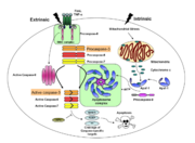
Any apoptotic signal received by the cell causes the activation of initiator caspases (-8 and -9) by associating with another protein platform to form a functional holoenzyme. These initiator caspases then cleaves the executioner caspases -3, -6, -7. Caspase-3 specifically functions to cleave both caspase-6 and -7, which in turn cleave their respective targets to induce cell death. Aside from being able to activate caspase-6 and -7, caspase-3 also regulates caspase-9 activity, operating via feedback loop. These dual action of caspase-3 confers its distinct regulatory mechanisms, resulting a wider extent of effects in the apoptotic cascade.
| |||||||||||
Caspase-3 Loop Bundle and Active Site
| |||||||||||
Caspase-3 Regulation
| |||||||||||
Proteopedia Page Contributors and Editors (what is this?)
Scott Eron, Banyuhay P. Serrano, Alexander Berchansky, Yunlong Zhao, Jaime Prilusky, Michal Harel


