Sandbox Reserved 716
From Proteopedia
| Line 26: | Line 26: | ||
== '''3D structure''' == | == '''3D structure''' == | ||
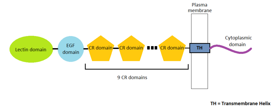
| - | P-Selectin is a protein composed by 162 amino-acids residues in 4 different chains A, B, C and D. There are many domains in this protein : the '''EGF domain''', the '''lectin domain''' and | + | P-Selectin is a protein composed by 162 amino-acids residues in 4 different chains A, B, C and D. There are many domains in this protein. Three domains represent the extracellular part of the protein : the '''EGF domain''', the '''lectin domain''' and nine '''consensus repeat (CR) domains'''. Finally, we find a transmembrane helix and a cytoplasmic domain. |
[[Image:Selectin_Structure.png|900px|center]] | [[Image:Selectin_Structure.png|900px|center]] | ||
| - | EGF domain (Epidermal Growth Factor) coutains 30 to 40 amino-acids residues | + | [[Image:PDB_1hre_EBI.jpg|200px|right|thumb| EGF domain]] EGF domain (Epidermal Growth Factor) coutains 30 to 40 amino-acids residues. This domain is composed by 6 cysteine residues which can form 3 disulfide bonds. EGF domain is formed with two-stranded β-sheet followed by a loop to a short C-terminal two-stranded β-sheet. These two β-sheets are usually denoted as the major (N-terminal) and minor (C-terminal) sheets. |
| - | [[Image: | + | |
| + | |||
| + | |||
| + | |||
| + | |||
| + | |||
| + | |||
| + | |||
| + | |||
| + | |||
| + | |||
| + | |||
| + | |||
| + | |||
| + | [[Image:368px-Pselectin.PNG|200px|left|thumb| P selectin lectin bound to sugar]] Lectin domains (also known as C-type lectin domains) are classified in 17 groups (from I to XVII). P-Selectin lectin bellow to the group IV. | ||
| + | |||
| + | |||
| + | |||
| + | |||
| + | |||
| + | |||
| + | |||
| + | |||
| + | |||
| + | |||
| + | |||
| + | |||
| + | |||
| + | |||
| + | |||
| + | |||
| + | |||
| + | |||
| + | |||
| + | |||
| + | |||
| + | |||
| + | |||
| + | |||
| + | |||
| + | |||
| + | |||
| + | |||
| - | Lectin domains (also known as C-type lectin domains) are classified in 17 groups (from I to XVII). P-Selectin lectin bellow to the group IV. | ||
| - | [[Image:368px-Pselectin.PNG|200px|center|thumb| P selectin lectin bound to sugar]] | ||
Revision as of 12:37, 3 January 2013
|
Crystal structure of P-selectin lectin/EGF domains complexed with SLeX
Contents |
Introduction
Selectins are proteins that are include in a family of cell adhesion receptor involved in the leukocyte extravasation. There are 3 kinds of selectins :
E selectin localized in endothelial cells, L selectin found in leukocytes, and P selectins in platelets and endothelial cells.
In this page we will be focused only on P-Selectin.
3D structure
P-Selectin is a protein composed by 162 amino-acids residues in 4 different chains A, B, C and D. There are many domains in this protein. Three domains represent the extracellular part of the protein : the EGF domain, the lectin domain and nine consensus repeat (CR) domains. Finally, we find a transmembrane helix and a cytoplasmic domain.
Different role of the P-selectin
Role in leukocyte extravasation
Leukocyte extravasation is the movement of leukocytes out of the circulatory system. First, the leukocyte is attracted by cytokines, secreted near the site of infection. Then, this leukocyte slows down and begin rolling along the surface of the vessel. He binds then tightly the vessel and immobilizates himself. Finally, he passes through gaps between epithelial cells. By this mecanism, the leukocyte arrives on the site of infection to neutralize the infection agent.
Role in platelets recruitment
Role in cancer
External resources
Protein Data Bank file 1G1R
References
1. http://cro.sagepub.com/content/10/3/337.full.pdf
2. Somers WS, Tang J, Shaw GD, Camphausen RT. Insights into the molecular basis of leukocyte tethering and rolling revealed by structures of P- and E-selectin bound to SLe(X) and PSGL-1. Cell. 2000 Oct 27;103(3):467-79. PMID : 11081633
Proteopedia Page Contributors and Editors
Delphine Trelat, Cécile Ehrhardt

