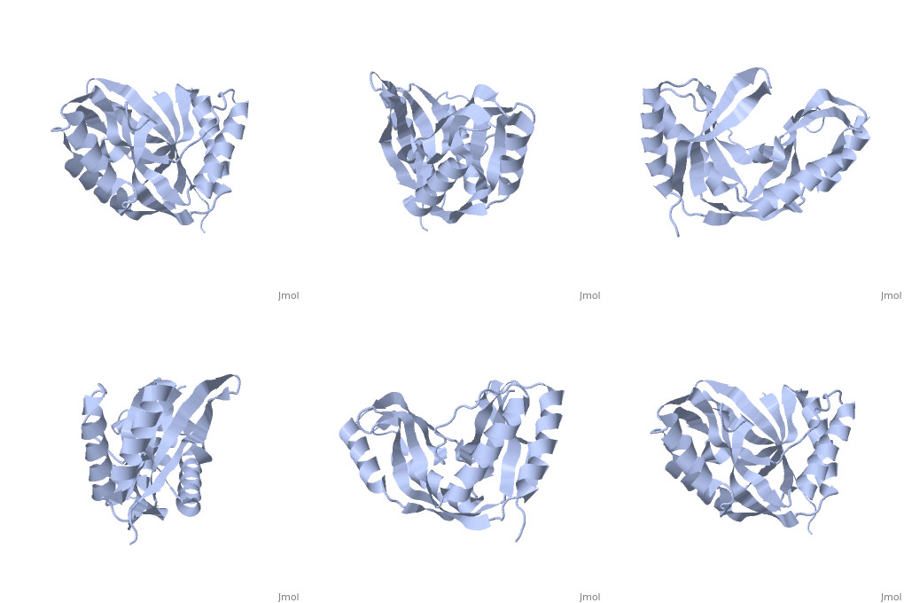
1vgj
From Proteopedia
(New page: 200px<br /><applet load="1vgj" size="450" color="white" frame="true" align="right" spinBox="true" caption="1vgj, resolution 1.94Å" /> '''Crystal structure of...) |
|||
| Line 1: | Line 1: | ||
| - | [[Image:1vgj.gif|left|200px]]<br /><applet load="1vgj" size=" | + | [[Image:1vgj.gif|left|200px]]<br /><applet load="1vgj" size="350" color="white" frame="true" align="right" spinBox="true" |
caption="1vgj, resolution 1.94Å" /> | caption="1vgj, resolution 1.94Å" /> | ||
'''Crystal structure of 2'-5' RNA ligase from Pyrococcus horikoshii'''<br /> | '''Crystal structure of 2'-5' RNA ligase from Pyrococcus horikoshii'''<br /> | ||
==Overview== | ==Overview== | ||
| - | Bacterial and archaeal 2'-5' RNA ligases, members of the 2H | + | Bacterial and archaeal 2'-5' RNA ligases, members of the 2H phosphoesterase superfamily, catalyze the linkage of the 5' and 3' exons via a 2'-5'-phosphodiester bond during tRNA-precursor splicing. The crystal structure of the 2'-5' RNA ligase PH0099 from Pyrococcus horikoshii OT3 was solved at 1.94 A resolution (PDB code 1vgj). The molecule has a bilobal alpha+beta arrangement with two antiparallel beta-sheets constituting a V-shaped active-site cleft, as found in other members of the 2H phosphoesterase superfamily. The present structure was significantly different from that determined previously at 2.4 A resolution (PDB code 1vdx) in the active-site cleft; the entrance to the cleft is wider and the active site is easily accessible to the substrate (RNA precursor) in our structure. Structural comparison with the 2'-5' RNA ligase from Thermus thermophilus HB8 also revealed differences in the RNA precursor-binding region. The structural differences in the active-site residues (tetrapeptide motifs H-X-T/S-X) between the members of the 2H phosphoesterase superfamily are discussed. |
==About this Structure== | ==About this Structure== | ||
| - | 1VGJ is a [http://en.wikipedia.org/wiki/Single_protein Single protein] structure of sequence from [http://en.wikipedia.org/wiki/Pyrococcus_horikoshii Pyrococcus horikoshii]. Full crystallographic information is available from [http:// | + | 1VGJ is a [http://en.wikipedia.org/wiki/Single_protein Single protein] structure of sequence from [http://en.wikipedia.org/wiki/Pyrococcus_horikoshii Pyrococcus horikoshii]. Full crystallographic information is available from [http://oca.weizmann.ac.il/oca-bin/ocashort?id=1VGJ OCA]. |
==Reference== | ==Reference== | ||
| - | The structure of Pyrococcus horikoshii 2'-5' RNA ligase at 1.94 A resolution reveals a possible open form with a wider active-site cleft., Gao YG, Yao M, Okada A, Tanaka I, Acta | + | The structure of Pyrococcus horikoshii 2'-5' RNA ligase at 1.94 A resolution reveals a possible open form with a wider active-site cleft., Gao YG, Yao M, Okada A, Tanaka I, Acta Crystallogr Sect F Struct Biol Cryst Commun. 2006 Dec 1;62(Pt, 12):1196-200. Epub 2006 Nov 30. PMID:[http://ispc.weizmann.ac.il//pmbin/getpm?pmid=17142895 17142895] |
[[Category: Pyrococcus horikoshii]] | [[Category: Pyrococcus horikoshii]] | ||
[[Category: Single protein]] | [[Category: Single protein]] | ||
| Line 21: | Line 21: | ||
[[Category: structural genomics]] | [[Category: structural genomics]] | ||
| - | ''Page seeded by [http:// | + | ''Page seeded by [http://oca.weizmann.ac.il/oca OCA ] on Thu Feb 21 15:35:02 2008'' |
Revision as of 13:35, 21 February 2008
|
Crystal structure of 2'-5' RNA ligase from Pyrococcus horikoshii
Overview
Bacterial and archaeal 2'-5' RNA ligases, members of the 2H phosphoesterase superfamily, catalyze the linkage of the 5' and 3' exons via a 2'-5'-phosphodiester bond during tRNA-precursor splicing. The crystal structure of the 2'-5' RNA ligase PH0099 from Pyrococcus horikoshii OT3 was solved at 1.94 A resolution (PDB code 1vgj). The molecule has a bilobal alpha+beta arrangement with two antiparallel beta-sheets constituting a V-shaped active-site cleft, as found in other members of the 2H phosphoesterase superfamily. The present structure was significantly different from that determined previously at 2.4 A resolution (PDB code 1vdx) in the active-site cleft; the entrance to the cleft is wider and the active site is easily accessible to the substrate (RNA precursor) in our structure. Structural comparison with the 2'-5' RNA ligase from Thermus thermophilus HB8 also revealed differences in the RNA precursor-binding region. The structural differences in the active-site residues (tetrapeptide motifs H-X-T/S-X) between the members of the 2H phosphoesterase superfamily are discussed.
About this Structure
1VGJ is a Single protein structure of sequence from Pyrococcus horikoshii. Full crystallographic information is available from OCA.
Reference
The structure of Pyrococcus horikoshii 2'-5' RNA ligase at 1.94 A resolution reveals a possible open form with a wider active-site cleft., Gao YG, Yao M, Okada A, Tanaka I, Acta Crystallogr Sect F Struct Biol Cryst Commun. 2006 Dec 1;62(Pt, 12):1196-200. Epub 2006 Nov 30. PMID:17142895
Page seeded by OCA on Thu Feb 21 15:35:02 2008

