This old version of Proteopedia is provided for student assignments while the new version is undergoing repairs. Content and edits done in this old version of Proteopedia after March 1, 2026 will eventually be lost when it is retired in about June of 2026.
Apply for new accounts at the new Proteopedia. Your logins will work in both the old and new versions.
1x0p
From Proteopedia
(New page: 200px<br /><applet load="1x0p" size="450" color="white" frame="true" align="right" spinBox="true" caption="1x0p, resolution 2.0Å" /> '''Structure of a cyanob...) |
|||
| Line 1: | Line 1: | ||
| - | [[Image:1x0p.gif|left|200px]]<br /><applet load="1x0p" size=" | + | [[Image:1x0p.gif|left|200px]]<br /><applet load="1x0p" size="350" color="white" frame="true" align="right" spinBox="true" |
caption="1x0p, resolution 2.0Å" /> | caption="1x0p, resolution 2.0Å" /> | ||
'''Structure of a cyanobacterial BLUF protein, Tll0078'''<br /> | '''Structure of a cyanobacterial BLUF protein, Tll0078'''<br /> | ||
==Overview== | ==Overview== | ||
| - | The sensor proteins for blue light using the FAD (BLUF) domain belong to | + | The sensor proteins for blue light using the FAD (BLUF) domain belong to the third family of the photoreceptor proteins using a flavin chromophore, where the other two families are phototropins and cryptochromes. As the first structure of this BLUF domain, we have determined the crystal structure of the Tll0078 protein from Thermosynechococcus elongatus BP-1, which contains a BLUF domain bound to FAD, at 2A resolution. Five Tll0078 monomers are located around the non-crystallographic 5-fold axis to form a pentamer, and two pentamers related by 2-fold non-crystallographic symmetry form a decameric assembly. The monomer consists of two domains, the BLUF domain at the N-terminal region and the C-terminal domain. The overall structure of the BLUF domain consists of a five-stranded mixed beta-sheet with two alpha-helices running parallel with it. The isoalloxazine ring of FAD is accommodated in a pocket formed by several highly conserved amino acid residues in the BLUF domain. Of these, the three apparent key residues (Asn31, Asn32 and Gln50) were substituted with Ala. Mutant proteins of N31A and N32A showed a nearly normal 10nm spectral shift of the flavin upon illumination, while the Q50A mutant did not exhibit such a shift at all. On the basis of the crystal structure, we discussed a possible role of Gln50, which is structurally and functionally linked with the critical Tyr8 (FAD-Gln50-Tyr8 network), with regard to the light-induced spectral shift of the BLUF proteins. |
==About this Structure== | ==About this Structure== | ||
| - | 1X0P is a [http://en.wikipedia.org/wiki/Single_protein Single protein] structure of sequence from [http://en.wikipedia.org/wiki/Thermosynechococcus_elongatus Thermosynechococcus elongatus] with FAD as [http://en.wikipedia.org/wiki/ligand ligand]. Full crystallographic information is available from [http:// | + | 1X0P is a [http://en.wikipedia.org/wiki/Single_protein Single protein] structure of sequence from [http://en.wikipedia.org/wiki/Thermosynechococcus_elongatus Thermosynechococcus elongatus] with <scene name='pdbligand=FAD:'>FAD</scene> as [http://en.wikipedia.org/wiki/ligand ligand]. Full crystallographic information is available from [http://oca.weizmann.ac.il/oca-bin/ocashort?id=1X0P OCA]. |
==Reference== | ==Reference== | ||
| Line 24: | Line 24: | ||
[[Category: tll0078]] | [[Category: tll0078]] | ||
| - | ''Page seeded by [http:// | + | ''Page seeded by [http://oca.weizmann.ac.il/oca OCA ] on Thu Feb 21 15:49:57 2008'' |
Revision as of 13:49, 21 February 2008
|
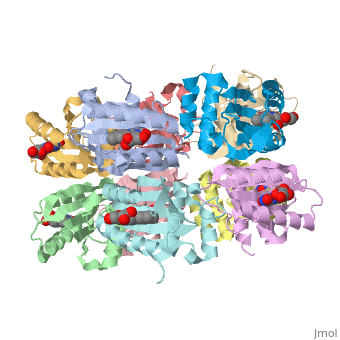
Structure of a cyanobacterial BLUF protein, Tll0078
Overview
The sensor proteins for blue light using the FAD (BLUF) domain belong to the third family of the photoreceptor proteins using a flavin chromophore, where the other two families are phototropins and cryptochromes. As the first structure of this BLUF domain, we have determined the crystal structure of the Tll0078 protein from Thermosynechococcus elongatus BP-1, which contains a BLUF domain bound to FAD, at 2A resolution. Five Tll0078 monomers are located around the non-crystallographic 5-fold axis to form a pentamer, and two pentamers related by 2-fold non-crystallographic symmetry form a decameric assembly. The monomer consists of two domains, the BLUF domain at the N-terminal region and the C-terminal domain. The overall structure of the BLUF domain consists of a five-stranded mixed beta-sheet with two alpha-helices running parallel with it. The isoalloxazine ring of FAD is accommodated in a pocket formed by several highly conserved amino acid residues in the BLUF domain. Of these, the three apparent key residues (Asn31, Asn32 and Gln50) were substituted with Ala. Mutant proteins of N31A and N32A showed a nearly normal 10nm spectral shift of the flavin upon illumination, while the Q50A mutant did not exhibit such a shift at all. On the basis of the crystal structure, we discussed a possible role of Gln50, which is structurally and functionally linked with the critical Tyr8 (FAD-Gln50-Tyr8 network), with regard to the light-induced spectral shift of the BLUF proteins.
About this Structure
1X0P is a Single protein structure of sequence from Thermosynechococcus elongatus with as ligand. Full crystallographic information is available from OCA.
Reference
Structure of a cyanobacterial BLUF protein, Tll0078, containing a novel FAD-binding blue light sensor domain., Kita A, Okajima K, Morimoto Y, Ikeuchi M, Miki K, J Mol Biol. 2005 May 27;349(1):1-9. Epub 2005 Apr 9. PMID:15876364
Page seeded by OCA on Thu Feb 21 15:49:57 2008
Categories: Single protein | Thermosynechococcus elongatus | Ikeuchi, M. | Kita, A. | Miki, K. | Morimoto, Y. | Okajima, K. | FAD | Bluf | Fad | Structural genomics | Tll0078
