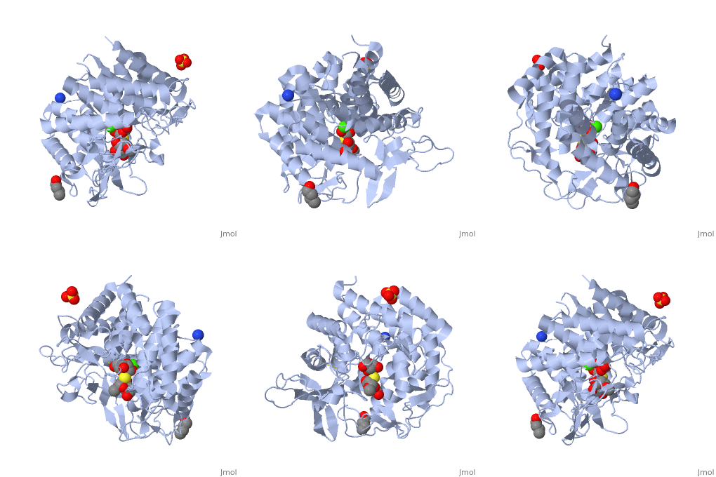
1x9d
From Proteopedia
(New page: 200px<br /> <applet load="1x9d" size="450" color="white" frame="true" align="right" spinBox="true" caption="1x9d, resolution 1.410Å" /> '''Crystal Structure ...) |
|||
| Line 1: | Line 1: | ||
| - | [[Image:1x9d.gif|left|200px]]<br /> | + | [[Image:1x9d.gif|left|200px]]<br /><applet load="1x9d" size="350" color="white" frame="true" align="right" spinBox="true" |
| - | <applet load="1x9d" size=" | + | |
caption="1x9d, resolution 1.410Å" /> | caption="1x9d, resolution 1.410Å" /> | ||
'''Crystal Structure Of Human Class I alpha-1,2-Mannosidase In Complex With Thio-Disaccharide Substrate Analogue'''<br /> | '''Crystal Structure Of Human Class I alpha-1,2-Mannosidase In Complex With Thio-Disaccharide Substrate Analogue'''<br /> | ||
==Overview== | ==Overview== | ||
| - | Quality control in the endoplasmic reticulum (ER) determines the fate of | + | Quality control in the endoplasmic reticulum (ER) determines the fate of newly synthesized glycoproteins toward either correct folding or disposal by ER-associated degradation. Initiation of the disposal process involves selective trimming of N-glycans attached to misfolded glycoproteins by ER alpha-mannosidase I and subsequent recognition by the ER degradation-enhancing alpha-mannosidase-like protein family of lectins, both members of glycosylhydrolase family 47. The unusual inverting hydrolytic mechanism catalyzed by members of this family is investigated here by a combination of kinetic and binding analyses of wild type and mutant forms of human ER alpha-mannosidase I as well as by structural analysis of a co-complex with an uncleaved thiodisaccharide substrate analog. These data reveal the roles of potential catalytic acid and base residues and the identification of a novel (3)S(1) sugar conformation for the bound substrate analog. The co-crystal structure described here, in combination with the (1)C(4) conformation of a previously identified co-complex with the glycone mimic, 1-deoxymannojirimycin, indicates that glycoside bond cleavage proceeds through a least motion conformational twist of a properly predisposed substrate in the -1 subsite. A novel (3)H(4) conformation is proposed as the exploded transition state. |
==About this Structure== | ==About this Structure== | ||
| - | 1X9D is a [http://en.wikipedia.org/wiki/Single_protein Single protein] structure of sequence from [http://en.wikipedia.org/wiki/Homo_sapiens Homo sapiens] with CA, SO4, SMD and BU1 as [http://en.wikipedia.org/wiki/ligands ligands]. Active as [http://en.wikipedia.org/wiki/Mannosyl-oligosaccharide_1,2-alpha-mannosidase Mannosyl-oligosaccharide 1,2-alpha-mannosidase], with EC number [http://www.brenda-enzymes.info/php/result_flat.php4?ecno=3.2.1.113 3.2.1.113] Full crystallographic information is available from [http:// | + | 1X9D is a [http://en.wikipedia.org/wiki/Single_protein Single protein] structure of sequence from [http://en.wikipedia.org/wiki/Homo_sapiens Homo sapiens] with <scene name='pdbligand=CA:'>CA</scene>, <scene name='pdbligand=SO4:'>SO4</scene>, <scene name='pdbligand=SMD:'>SMD</scene> and <scene name='pdbligand=BU1:'>BU1</scene> as [http://en.wikipedia.org/wiki/ligands ligands]. Active as [http://en.wikipedia.org/wiki/Mannosyl-oligosaccharide_1,2-alpha-mannosidase Mannosyl-oligosaccharide 1,2-alpha-mannosidase], with EC number [http://www.brenda-enzymes.info/php/result_flat.php4?ecno=3.2.1.113 3.2.1.113] Full crystallographic information is available from [http://oca.weizmann.ac.il/oca-bin/ocashort?id=1X9D OCA]. |
==Reference== | ==Reference== | ||
| Line 16: | Line 15: | ||
[[Category: Single protein]] | [[Category: Single protein]] | ||
[[Category: Karaveg, K.]] | [[Category: Karaveg, K.]] | ||
| - | [[Category: Liu, Z | + | [[Category: Liu, Z J.]] |
| - | [[Category: Moremen, K | + | [[Category: Moremen, K W.]] |
[[Category: Siriwardena, A.]] | [[Category: Siriwardena, A.]] | ||
[[Category: Tempel, W.]] | [[Category: Tempel, W.]] | ||
| - | [[Category: Wang, B | + | [[Category: Wang, B C.]] |
[[Category: BU1]] | [[Category: BU1]] | ||
[[Category: CA]] | [[Category: CA]] | ||
| Line 29: | Line 28: | ||
[[Category: substrate analogue]] | [[Category: substrate analogue]] | ||
| - | ''Page seeded by [http:// | + | ''Page seeded by [http://oca.weizmann.ac.il/oca OCA ] on Thu Feb 21 15:52:22 2008'' |
Revision as of 13:52, 21 February 2008
|
Crystal Structure Of Human Class I alpha-1,2-Mannosidase In Complex With Thio-Disaccharide Substrate Analogue
Overview
Quality control in the endoplasmic reticulum (ER) determines the fate of newly synthesized glycoproteins toward either correct folding or disposal by ER-associated degradation. Initiation of the disposal process involves selective trimming of N-glycans attached to misfolded glycoproteins by ER alpha-mannosidase I and subsequent recognition by the ER degradation-enhancing alpha-mannosidase-like protein family of lectins, both members of glycosylhydrolase family 47. The unusual inverting hydrolytic mechanism catalyzed by members of this family is investigated here by a combination of kinetic and binding analyses of wild type and mutant forms of human ER alpha-mannosidase I as well as by structural analysis of a co-complex with an uncleaved thiodisaccharide substrate analog. These data reveal the roles of potential catalytic acid and base residues and the identification of a novel (3)S(1) sugar conformation for the bound substrate analog. The co-crystal structure described here, in combination with the (1)C(4) conformation of a previously identified co-complex with the glycone mimic, 1-deoxymannojirimycin, indicates that glycoside bond cleavage proceeds through a least motion conformational twist of a properly predisposed substrate in the -1 subsite. A novel (3)H(4) conformation is proposed as the exploded transition state.
About this Structure
1X9D is a Single protein structure of sequence from Homo sapiens with , , and as ligands. Active as Mannosyl-oligosaccharide 1,2-alpha-mannosidase, with EC number 3.2.1.113 Full crystallographic information is available from OCA.
Reference
Mechanism of class 1 (glycosylhydrolase family 47) {alpha}-mannosidases involved in N-glycan processing and endoplasmic reticulum quality control., Karaveg K, Siriwardena A, Tempel W, Liu ZJ, Glushka J, Wang BC, Moremen KW, J Biol Chem. 2005 Apr 22;280(16):16197-207. Epub 2005 Feb 15. PMID:15713668
Page seeded by OCA on Thu Feb 21 15:52:22 2008

