This old version of Proteopedia is provided for student assignments while the new version is undergoing repairs. Content and edits done in this old version of Proteopedia after March 1, 2026 will eventually be lost when it is retired in about June of 2026.
Apply for new accounts at the new Proteopedia. Your logins will work in both the old and new versions.
1dtg
From Proteopedia
(Difference between revisions)
| Line 1: | Line 1: | ||
| - | [[Image:1dtg.png|left|200px]] | ||
| - | |||
{{STRUCTURE_1dtg| PDB=1dtg | SCENE= }} | {{STRUCTURE_1dtg| PDB=1dtg | SCENE= }} | ||
| - | |||
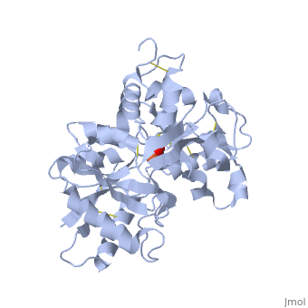
===HUMAN TRANSFERRIN N-LOBE MUTANT H249E=== | ===HUMAN TRANSFERRIN N-LOBE MUTANT H249E=== | ||
| - | |||
{{ABSTRACT_PUBMED_10684598}} | {{ABSTRACT_PUBMED_10684598}} | ||
Revision as of 09:16, 11 March 2013
| |||||||||
| 1dtg, resolution 2.40Å () | |||||||||
|---|---|---|---|---|---|---|---|---|---|
| Ligands: | , | ||||||||
| |||||||||
| |||||||||
| Resources: | FirstGlance, OCA, RCSB, PDBsum | ||||||||
| Coordinates: | save as pdb, mmCIF, xml | ||||||||

Contents |
HUMAN TRANSFERRIN N-LOBE MUTANT H249E
Template:ABSTRACT PUBMED 10684598
About this Structure
1dtg is a 1 chain structure with sequence from Homo sapiens. Full crystallographic information is available from OCA.
See Also
- Cartoon backbone representation
- Secondary structure
- Sheets in Proteins
- Spacefilling representation
- Transferrin
Reference
- MacGillivray RT, Bewley MC, Smith CA, He QY, Mason AB, Woodworth RC, Baker EN. Mutation of the iron ligand His 249 to Glu in the N-lobe of human transferrin abolishes the dilysine "trigger" but does not significantly affect iron release. Biochemistry. 2000 Feb 15;39(6):1211-6. PMID:10684598
