Sandbox Reserved 696
From Proteopedia
| Line 22: | Line 22: | ||
<Structure load='3KG2' size='500' frame='true' align='right' caption='Insert caption here' scene='Glutamate Receptor' /> | <Structure load='3KG2' size='500' frame='true' align='right' caption='Insert caption here' scene='Glutamate Receptor' /> | ||
| + | |||
| + | <Structure load='1PBQ' size='500' frame='true' align='right' caption='Insert caption here' scene='ligand binding core in complex with DCKA' /> | ||
| + | |||
| + | <Structure load='2CMO' size='500' frame='true' align='right' caption='Insert caption here' scene='ligand binding core dimer in complex with glutamate and antagonist' /> | ||
Revision as of 02:29, 1 May 2013
| This Sandbox is Reserved from 30/01/2013, through 30/12/2013 for use in the course "Biochemistry II" taught by Hannah Tims at the Messiah College. This reservation includes Sandbox Reserved 686 through Sandbox Reserved 700. |
To get started:
More help: Help:Editing |
Contents |
Background
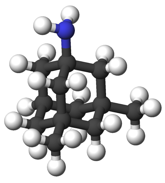
Memantine aka ... memantine is a key alzheimers drug that helps improve memory and learning inability Doses of Memantine llows normal physiological activity of glutamate (NMDAR) receptors but prevents overstimulation. Overstimulation causes excitotoxicity. (what it is and how it is believed to attribute to Alzheimer's.)
NMDAR Structure
general structure subunits (insert picture of NR1 and NR2)
|
interactions between subunits (show interactions)
|
|
|
NMDA receptor is heterotetramer of two GluN1 (or NR1) and two GluN2 (or NR2) subunits. Each subunit acts as a distinct functional unit. The extracellular domain contains two globular structures, a modulating domain and a ligand binding domain. NR1 bind co-agonists and NR2 binds glutamate. Agonist binding module links to a membrane domain, which has three transmembrane segments and a re-entrant loop. The membrane domain contributes residues to the channel pore. The cytoplasmic domain contains residues that can be modified by series of protein kinases and phosphatases. The cytoplasmic domain also has residues that interact with scaffolding proteins (Vance, 1-2).
NMDAR Binding Sites
places for ligand, agonist, antagonist (show binding spots) interactions by which they bind (show aa interactions) which subunit they bind to
NMDAR Ion Channel
agonist bind, then conformational change, then ion flux in (show binding spot in receptor) (show channel)
Memantine Uses
recreational drugs could potentially help any disease that is subject to excitotoxicity like Parkinson, Alzheimers, Etc.
GluA2 Structure
AMPA IGluRs form homotetramers composed of distal subunit partners and proximal subunits partners. Each subunit includes an extracellular amino terminal domain (ATD) which is responsible for receptor trafficking within the membrane, a ligand-binding domain (LBD) which activates the receptor upon binding glutamate, and a transmembrane domain (TMD) which forms the membrane-spanning ion channel. Also present is a carboxy-terminal domain involved in receptor localization and regulation, although the structure of this domain has not been solved.[2] The structure of AMPA IGluRs or in this case GluA2, is unique in that the symmetry of the receptor changes depending on the domain. The ATD has a local two-fold symmetry, the LBD has a two-fold symmetry, while the TMD has a four-fold symmetry. Here is a morph depicting the differnce between subunit type A and B. This symmetry mismatch has implications for function of the receptor with subunits behaving differently depending upon their orientation despite identical primary sequence.[2] For an excellent analysis, see: Glutamate Receptor Symmetry Analysis
The Amino Terminal Domain
The ATD is responsible for receptor assembly, trafficking and localization. It has two unique sets of interactions which hold the tetramer together. The first set of interactions is present in each pair of dimers and involves both hydrogen bonding and hydrophobic interactions. The second set, which includes residues Ile 203, Thr 204, Ile 205, and Val 209 on both chains among others, effectively holds the pair of dimers together at an angle that is roughly 24 degrees off of the overall two-fold axis.[2][7]
The Transmembrane Domain
The TMD has a pore structure that is nearly identical to that of the Potassium Channel. With complete four-fold symmetry, 16 helices form a precise pore through which cations can flow through. In the current, inhibitor bound structure, the M3 helices cross at a highly conserved SYTANLAAF motif, with Thr 617, Ala 621, and Thr 625 occluding the ion permeation pathway.[2] The narrowest part of the channel includes the residues Thr 625, Ala 621, and Thr 617, but does not distinguish between positive cations like in the Potassium Channel. Located next to this narrow region lies Alanine 622, which is replaced with a threonine in the Lurcher mouse model mentioned previously. This mutation, which introduces a much bulkier residue, prevents the helices from closing properly, resulting in a constitutively open ion channel.[2]
The Ligand Binding Domain
The LBD is located just above the TMD and has an overall two-fold axis of symmetry. Within each LBD lies the so-called “clamshell”. This structure is responsible for binding glutamate and “sensitizing” the receptor to allow passage of cations through the channel. Residues Pro 89, Leu 90, Arg 96, Ser 142, & Glu 193 among others (residue numbers in 1ftj model), which are responsible for tightly binding glutamate within the clamshell, are highly conserved. Glutamate binding causes a conformational change (Alternate View) in the LBD which pulls the M3 helices in the TMD apart, opening the channel and allowing for cation passage. A morph of the conformational change in the LBD upon glutamate binding can be seen here. Uniquely, due to the varied importance of the homotetramer subunits due to symmetry mismatch, the interaction of glutamate with the distal subunits is predicted to result in a greater conformational change. Thus these distal subunits play a more critical role in channel sensitization and activation.[2]

