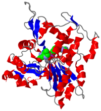
Crystal Structure of F-actin,
2zwh (
F-actin) units are also referred to as
microfilaments [1] and are highly conserved, proteinous components found near ubiquitously in eukaryotic cytoskeletons. F-actin and other
actin proteins generally have structural roles in cells.
Introduction
Actin is found in nearly all eukaryotic cells and is known primarily for its function as a structural and translocation protein. It also has an ATPase function, as it hydrolyzes ATP to ADP and Pi and undergoes conformational changes with each hydrolysis. Actin belongs to the actin superfamily, which includes other proteins such as Hsp70(DnaK), Hsc70, and hexokinase, because of its nucleotide-dependent conformational change[2]. Because of the similarity observed in Escherichia Coli's, Hsc70 and ATPase domain of actin, it is believed that the two proteins have a common ancestry[3]. Prokaryotes are not known to have actin, but do however have an actin homologue, MreB, which also leads to the idea of possible common ancestory[4].
Actin occurs in two forms: globular actin (G-actin), the free monomeric units of actin, and filamentous actin (F-actin) which is the polymer form. These two forms exist in a dynamic equilibrium with one another as ATP-associated polymerization and depolymerization occur continuously within the cell. The monomer units in the F-actin possess a form that is distinct from the free monomeric form and it is a result of that change that the more specific ATPase activity may be observed.
Assembly
(1J6Z).
G-actin is the free monomeric form of actin which polymerizes to F-actin. The structures of globular and filamentous actin are distinct from one another in numerous ways, despite the fact that G-actin comprises F-actin. When the monomeric actin becomes polymerized into F-actin, the unit becomes flattened. Also, F-actin possesses an ATPase function which is minimal in G-actin. The domains and active site are the same in terms of constituent components and will be discussed later in terms of the F-actin monomer.
G-actin appears to have more ligands in its structure, external to the active site. Only 3 of the 5 are believed to actually exist in solution and are believed to contribute to the polymerization of G-actin to F-actin[5]. This representation of G-actin also possesses an which is observed in some actin crystalline structures but not necessarily[5]. The observed molecule on Cys374, was used to block polymerization activity so the crystal of G-actin could be observed[5]
Formation of F-actin is a dynamic process of assembly and disassembly which has been termed “treadmilling”.
The transition between G and F-actin begins with a stabilized oligomer of ATP-actin units formed through a nucleation-condensation type fold pattern[6]. Addition of ATP-monomeric units to either end subsequently occurs, however, because of a difference in charge polarity in the two ends, there is preferential addition to what is termed the "plus (+) end" or the "barbed-end". On the opposite end, the "minus (-) end" or the "pointed end", there is preferential dissociation of actin units[7].
After attachment of the ATP-bound actin, hydrolysis of the ATP occurs yielding the ADP and Pi bound state. Subsequent loss of a Pi leaves the ADP-actin state[8]. Because of the potential for addition or removal of monomeric units to occur at both ends, the assembly of F-actin may be described in terms of equilibrium. However, because the rate of ATP-actin association is ten-fold that of ADP-actin dissociation, the f-actin has the appearance of moving forward, or "treadmilling"[9]. ADP-actin monomers dissociate at the minus end and become recycled to ATP-actin so polymerization at the plus end may occur once again.
Structure
History of the structure
The F-actin protein was discovered by Straub in 1942[10]. The structure was speculated based on a low-resolution x-ray crystallograph found in 1990 by Holmes et al. and over this time, the "Holmes model" was accepted[11]. In contrast, the G-actin structure has been determined independently over 30 times. A higher resolution F-actin model was only recently deposited in the PDB databank in December 2008 by Oda et al. [10].
F-actin Monomer and Polymer
(2zwh)
Monomer
Each F-actin monomeric unit has, as part of its tertiary structure, several loops that are important to its assembly to the polymeric F-actin. These loops undergo conformational changes based on the state of the bound nucleotide or they serve as regions for adjacent monomeric actin units to bind to. The acts as a "switch" for conformations, based on the bound nucleotide[12]. The DNAse I-binding loop residues (40-50) in addition to undergoing conformational changes that impact stability bind DNAse I enzymes and are speculated to keep a hold on the DNAse I[2]. The hydrophobic loop , spanning residues 264-273, and the , spanning residues 165-172, function as sites to which adjacent actin monomer D-loops may bind to[13]. A similar function is noted for the residues (374-375).
The F-actin molecule as shown here consists of 375 residues(43kDa) and two ligands, ADP and Ca2+. It has two major domains separated by a nucleotide-binding cleft[10]. Depending on the state of the bound nucleotide, the most stable conformation of F-actin changes. In its ATP and ADP + Pi nucleotide bound states, it has a closed binding cleft. In its ADP only bound state, it has a wider binding cleft[6]A characteristic trait of actin is that the domains remain twisted relative to one another, despite the nucleotide-state-dependent conformational changes[10].
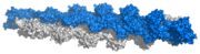
F-Actin polymer (based on Ken Holmes F-actin structure)
[3] Polymer
F-actin has the appearance of two right-handed helices, with a gradual twist around one another. It is actually composed of repeats of 13 actin units for every 6 left-handed turns, spanning a length of 350 Å[4].
Nucleotide-State-Dependent Conformational Changes
The state of the bound phosphorylated nucleotide affects what conformation the F-actin monomer undertakes. The presence of a gamma-phosphate in the active site causes the rotation of a Ser14 residue. This change leads to a methylated histidine (HIC73) becoming shifted, which alters the F-actin active site and causes a conformational change in the D-loop. The HIC73 is located in the "sensor loop", or the "switch" for linking changes in bound nucleotide to conformational changes[2]. In ATP-actin and ADP-Pi-actin, the D-loop is unstructured. In the ADP-bound form of F-actin, an alpha helix is commonly apparent in the D-loop of the monomer[6][2].
Although the alpha-helix is not observed in this Oda model of F-actin and is not seen in some other F-actin studies[10][14], it is acknowledged by Oda et. al that the experimental results could have lead to an extended alpha-helix in the model, as opposed to an extended disordered strand as the interacting segment between F-actin monomeric units[10].
Domains
(2zwh)
The structure of a single unit of F-actin arises from one polypeptide chain with two domains. The nucleotide binding cleft, site of ATP hydrolysis, can be observed between the two domains. Movement of the domains allows for the open and closed F-actin conformations.
Domain movement is made possible by rotation about the , shown in purple. According to Oda et al., during the transition from G- to F- actin, Domain 2 is believed to tilt 20° and fit itself with Domain 1, thus giving a flatter conformation than the free G-actin. It is not certain whether this flattening occurs before or after ATP hydrolysis[10]. Holmes[4] provides a simplified image of this domain movement and flattening[1].
Stability
The flattened folded form of F-actin requires different stabilization mechanisms than the free monomeric G-actin form. Stability of the F-actin complex is achieved by a series of involving arginine 206, 183, 177 (purple); glutamate 72(blue), aspartate 187(green), 179 and 4-methyl histidine 73(yellow). Additional stability is believed to arise from a break in the interaction between residues in the same half of their respective domains to a new interaction between where a much greater distance is observed between them[10].
Once the Pi is released, a conformational change on the D-loop results in the “softening” of the F-actin filament. That is, it makes the ADP-actin monomer more unstable and makes it more susceptible to cleavage [15]
Active Site
Upon actin binding on the plus end of the actin filament, the ATPase function is activated. The conformational change from G- to F- actin promotes the catalytic activity because of the 20° shift leading to a more closed binding site; this conformational change is stabilized also by the diagonal subdomain interaction between Leu110 and Thr194[10].
As a result of these conformational changes, the of actin is moved closer to the ATP-Ca2+ ligand. Gln137 holds a water molecule, and placing it in close proximity to ATP allows for cleavage of the gamma-phosphate. Release of the inorganic phosphate occurs via the conformational change of the flexible "D-loop" into an ordered alpha-helix (though not demonstrated by this model)[2].
Function
F-actin performs a structural, mechanical, and enzymatic role within eukaryotic cells. These functions are not necessarily exclusive of one another.
The dynamic functions of f-actin are heavily involved with cell migration[16].
F-actin is the most abundant component of the cytoskeleton of eukaryotes. It provides large amounts of tensile strength, considering its thin size. In cases where the flexibility is not desirable as a structural component, crosslinkages can be formed between F-actin polymers to give greater stiffness and support[7].
Elongation of F-actin branches leads to the phenomenon of pushing of the plasma membrane forward in lamellopodial and filopodial extension[8]. This process relies on the dynamic equilibrium state in which G- and F-actin exist, as it is the continual polymerization of actin units on the leading edge that propels the membrane extension. Without the enzymatic ATPase function of F-actin, this process would not be possible.
The relatively flatter shape of F-actin as compared to G-actin allows myosin to preferentially bind F-actin over G-actin. This means that F-actin, not G-actin, is the functional form of actin. It composes a large part of the thin filaments in conjunction with mysoin to give muscle contractions[4][17]. The structure of F-actin gives it large resistance to extensive forces, such as those experienced in muscle contraction[7].
3D structures of actin
Actin
References
- ↑ Microfilaments - Wikipedia, the free encyclopedia. http://en.wikipedia.org/wiki/Microfilament. Date accessed: March 16th, 2010.
- ↑ 2.0 2.1 2.2 2.3 2.4 Graceffa P, Dominguez R. Crystal structure of monomeric actin in the ATP state. Structural basis of nucleotide-dependent actin dynamics. J Biol Chem. 2003 Sep 5;278(36):34172-80. Epub 2003 Jun 17. PMID:12813032 doi:10.1074/jbc.M303689200
- ↑ 3.0 3.1 Holmes KC. Structural biology: actin in a twist. Nature. 2009 Jan 22;457(7228):389-90. PMID:19158779 doi:10.1038/457389a
- ↑ 4.0 4.1 4.2 4.3 Holmes KC, Popp D, Gebhard W, Kabsch W. Atomic model of the actin filament. Nature. 1990 Sep 6;347(6288):44-9. PMID:2395461 doi:http://dx.doi.org/10.1038/347044a0
- ↑ 5.0 5.1 5.2 Otterbein LR, Graceffa P, Dominguez R. The crystal structure of uncomplexed actin in the ADP state. Science. 2001 Jul 27;293(5530):708-11. PMID:11474115 doi:10.1126/science.1059700
- ↑ 6.0 6.1 6.2 Pfaendtner J, Branduardi D, Parrinello M, Pollard TD, Voth GA. Nucleotide-dependent conformational states of actin. Proc Natl Acad Sci U S A. 2009 Aug 4;106(31):12723-8. Epub 2009 Jul 20. PMID:19620726
- ↑ 7.0 7.1 7.2 Mitchison TJ. Compare and contrast actin filaments and microtubules. Mol Biol Cell. 1992 Dec;3(12):1309-15. PMID:1493331
- ↑ 8.0 8.1 Chen H, Bernstein BW, Bamburg JR. Regulating actin-filament dynamics in vivo. Trends Biochem Sci. 2000 Jan;25(1):19-23. PMID:10637608
- ↑ Carlier MF, Pantaloni D. Direct evidence for ADP-Pi-F-actin as the major intermediate in ATP-actin polymerization. Rate of dissociation of Pi from actin filaments. Biochemistry. 1986 Dec 2;25(24):7789-92. PMID:3801442
- ↑ 10.0 10.1 10.2 10.3 10.4 10.5 10.6 10.7 10.8 Oda T, Iwasa M, Aihara T, Maeda Y, Narita A. The nature of the globular- to fibrous-actin transition. Nature. 2009 Jan 22;457(7228):441-5. PMID:19158791 doi:10.1038/nature07685
- ↑ Holmes KC, Popp D, Gebhard W, Kabsch W. Atomic model of the actin filament. Nature. 1990 Sep 6;347(6288):44-9. PMID:2395461 doi:http://dx.doi.org/10.1038/347044a0
- ↑ Oztug Durer ZA, Diraviyam K, Sept D, Kudryashov DS, Reisler E. F-actin structure destabilization and DNase I binding loop: fluctuations mutational cross-linking and electron microscopy analysis of loop states and effects on F-actin. J Mol Biol. 2010 Jan 22;395(3):544-57. Epub 2009 Nov 6. PMID:19900461 doi:10.1016/j.jmb.2009.11.001
- ↑ Oztug Durer ZA, Diraviyam K, Sept D, Kudryashov DS, Reisler E. F-actin structure destabilization and DNase I binding loop: fluctuations mutational cross-linking and electron microscopy analysis of loop states and effects on F-actin. J Mol Biol. 2010 Jan 22;395(3):544-57. Epub 2009 Nov 6. PMID:19900461 doi:10.1016/j.jmb.2009.11.001
- ↑ Dalhaimer P, Pollard TD, Nolen BJ. Nucleotide-mediated conformational changes of monomeric actin and Arp3 studied by molecular dynamics simulations. J Mol Biol. 2008 Feb 8;376(1):166-83. Epub 2007 Nov 28. PMID:18155236 doi:10.1016/j.jmb.2007.11.068
- ↑ Pfaendtner J, Lyman E, Pollard TD, Voth GA. Structure and dynamics of the actin filament. J Mol Biol. 2010 Feb 19;396(2):252-63. Epub 2009 Nov 18. PMID:19931282 doi:10.1016/j.jmb.2009.11.034
- ↑ Stricker J, Falzone T, Gardel ML. Mechanics of the F-actin cytoskeleton. J Biomech. 2010 Jan 5;43(1):9-14. Epub 2009 Nov 13. PMID:19913792 doi:10.1016/j.jbiomech.2009.09.003
- ↑ Holmes KC, Angert I, Kull FJ, Jahn W, Schroder RR. Electron cryo-microscopy shows how strong binding of myosin to actin releases nucleotide. Nature. 2003 Sep 25;425(6956):423-7. PMID:14508495 doi:10.1038/nature02005

