This old version of Proteopedia is provided for student assignments while the new version is undergoing repairs. Content and edits done in this old version of Proteopedia after March 1, 2026 will eventually be lost when it is retired in about June of 2026.
Apply for new accounts at the new Proteopedia. Your logins will work in both the old and new versions.
Single stranded binding protein
From Proteopedia
(Difference between revisions)
| Line 13: | Line 13: | ||
<StructureSection load='2vw9' size='500' side='right' frame='true' caption='Structure of Single Stranded DNA-Binding Protein bound to ssDNA (PDB entry [[2vw9]])' scene=''> | <StructureSection load='2vw9' size='500' side='right' frame='true' caption='Structure of Single Stranded DNA-Binding Protein bound to ssDNA (PDB entry [[2vw9]])' scene=''> | ||
| - | + | ||
==Structure== | ==Structure== | ||
| Line 36: | Line 36: | ||
</StructureSection> | </StructureSection> | ||
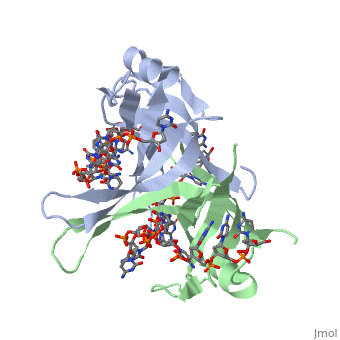
| + | <StructureSection load='1qvc' size='500' side='right' frame='true' caption='Structure of Single Stranded DNA-Binding Protein from ''E. coli'' (PDB entry [[1qvc]])' scene=''> | ||
| + | |||
| + | ==Binding Interactions between DNA and SSB of ''E. coli''== | ||
| + | Phe60 is an important DNA binding site. It has been shown to be the site for cross-linking. | ||
| + | Tryptophan and Lysine residues are important in binding as well. Treatments resulting in | ||
| + | modification of arginine, cysteine, or tyrosine residues had no effect on binding of SSB to | ||
| + | DNA, whereas modification of either lysine residues (with acetic anhydride) or tryptophan | ||
| + | residues (with N-bromosuccinimide) led to complete loss of binding activity <ref>PMID: 2087220</ref>. | ||
| + | The two tryptophan residues involved in DNA binding are Trp40 and Trp54, which was | ||
| + | determined by mutagenesis. One more binding site was determined by site-specific mutagenesis. | ||
| + | When His55 is substituted with Leu it decreases binding affinity. All of these residues | ||
| + | are found in a hydrophobic region, which is suitable for nucleotide base interactions. | ||
| + | </StructureSection> | ||
==See Also== | ==See Also== | ||
Revision as of 16:45, 1 November 2013
Sandbox Single Stranded DNA-Binding Protein (SSB)
| |||||||||||
| |||||||||||
| |||||||||||
See Also
References
- ↑ Meyer RR, Laine PS. The single-stranded DNA-binding protein of Escherichia coli. Microbiol Rev. 1990 Dec;54(4):342-80. PMID:2087220
- ↑ Meyer RR, Laine PS. The single-stranded DNA-binding protein of Escherichia coli. Microbiol Rev. 1990 Dec;54(4):342-80. PMID:2087220
- ↑ Meyer RR, Laine PS. The single-stranded DNA-binding protein of Escherichia coli. Microbiol Rev. 1990 Dec;54(4):342-80. PMID:2087220
Proteopedia Page Contributors and Editors (what is this?)
Refayat Ahsen, Rachel Craig, Michal Harel, Alexander Berchansky
