ATP-dependent DNA ligase from bacteriophage T7
From Proteopedia
(Difference between revisions)
(→ATP-DEPENDENT DNA LIGASE FROM BACTERIOPHAGE T7) |
|||
| Line 1: | Line 1: | ||
===ATP-DEPENDENT DNA LIGASE FROM BACTERIOPHAGE T7=== | ===ATP-DEPENDENT DNA LIGASE FROM BACTERIOPHAGE T7=== | ||
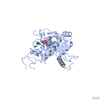
<StructureSection load='1a0i' size='350' side='right' caption='Structure of Bacteriophage T7 DNA Ligase(PDB entry [[1a0i]])' scene=''> | <StructureSection load='1a0i' size='350' side='right' caption='Structure of Bacteriophage T7 DNA Ligase(PDB entry [[1a0i]])' scene=''> | ||
| - | + | ==Overview (General Function)== | |
ATP-dependent DNA ligase from bacteriophage T7 (Caudovirales Podoviridae) is used to catalyze a phosphodiester bond between single-strand nicks in double-stranded DNA. This occurs in replication (connecting okazaki fragments), DNA repair (excision repair), and recombination. DNA ligases require either ATP (eukaryotes and viruses) or NAD+ (prokaryotes) as a cofactor. All ligases require a divalent cation for function. Bacteriophage T7 DNA ligase uses Magnesium in vivo. A range of pH 7.2-7.7 is ideal for enzymatic activity. T7 ligase has a molecular weight of 41 kDa. | ATP-dependent DNA ligase from bacteriophage T7 (Caudovirales Podoviridae) is used to catalyze a phosphodiester bond between single-strand nicks in double-stranded DNA. This occurs in replication (connecting okazaki fragments), DNA repair (excision repair), and recombination. DNA ligases require either ATP (eukaryotes and viruses) or NAD+ (prokaryotes) as a cofactor. All ligases require a divalent cation for function. Bacteriophage T7 DNA ligase uses Magnesium in vivo. A range of pH 7.2-7.7 is ideal for enzymatic activity. T7 ligase has a molecular weight of 41 kDa. | ||
| Line 8: | Line 8: | ||
| - | + | == Structure == | |
ATP-dependent DNA ligase from bacteriophage T7 is monomeric, forming a tertiary structure consisting of two domains (domain 1 and domain 2). Domain 1 (residues 2:240) contains the ATP binding site. Domain 1 is composed of six alpha helices which surround three antiparallel Beta sheets. Domain 2 (residues 241:349) is composed of an antiparallel Beta sheet and an alpha helix. A groove is formed between the two domains; this groove allows ATP to bind with domain 1. The ribose ring of ATP forms hydrogen bonds with the side chains of <scene name='56/567310/Arg_39_arg_55_glu_93/1'>Arg-39, Arg-55, and Glu-93</scene>. <scene name='56/567310/Lys232_lys238_lys34/2'>Lys-232, Lys-238, and Lys-34</scene> (the catalytic residue) form hydrogen bonds with the three phosphoryl groups of ATP. The 6-amino group of the adenine ring creates hydrogen bonds with the main-chain carbonyl of Ile-33 and the side chain of Glu-32. This could account for the use of ATP rather than GTP. While consisting of 359 residues, residues 121-127, 307-316, and 350-359 are not easily deciphered from the crystalline structure, and are therefore left out of the diagram. Domain 1 contains the N terminus, while domain 2 contains the C terminus. Multiple N and C terminii are shown in the diagram due to the missing residues. | ATP-dependent DNA ligase from bacteriophage T7 is monomeric, forming a tertiary structure consisting of two domains (domain 1 and domain 2). Domain 1 (residues 2:240) contains the ATP binding site. Domain 1 is composed of six alpha helices which surround three antiparallel Beta sheets. Domain 2 (residues 241:349) is composed of an antiparallel Beta sheet and an alpha helix. A groove is formed between the two domains; this groove allows ATP to bind with domain 1. The ribose ring of ATP forms hydrogen bonds with the side chains of <scene name='56/567310/Arg_39_arg_55_glu_93/1'>Arg-39, Arg-55, and Glu-93</scene>. <scene name='56/567310/Lys232_lys238_lys34/2'>Lys-232, Lys-238, and Lys-34</scene> (the catalytic residue) form hydrogen bonds with the three phosphoryl groups of ATP. The 6-amino group of the adenine ring creates hydrogen bonds with the main-chain carbonyl of Ile-33 and the side chain of Glu-32. This could account for the use of ATP rather than GTP. While consisting of 359 residues, residues 121-127, 307-316, and 350-359 are not easily deciphered from the crystalline structure, and are therefore left out of the diagram. Domain 1 contains the N terminus, while domain 2 contains the C terminus. Multiple N and C terminii are shown in the diagram due to the missing residues. | ||
| Line 15: | Line 15: | ||
| - | + | ==Catalytic function with DNA== | |
ATP-dependent DNA ligase from bacteriophage T7 amends a fractured DNA strand through esterification of a 5'- phosphoryl to a 3'- hydroxyl group. This mechanism occurs with the aid of ATP in several steps. First, the ligase is activated through a <scene name='56/567310/Amp_complex/1'>phosphoramidate bond with a lysine residue</scene> in the active site (Lys 34). A pyrophosphate leaves and the enzyme-AMP complex is formed. Next, the AMP is transferred to the 5' phosphate group at the nick in the DNA. Finally, T7 ligase creates the phosphodiester bond between the 5' -phosphoryl and the 3' – hydroxyl group, with AMP being freed. All ATP-dependent DNA ligases contain a conserved amino acid sequence of KxDGxR. This includes the lysine residue which binds the ATP in the groove between the two domains. | ATP-dependent DNA ligase from bacteriophage T7 amends a fractured DNA strand through esterification of a 5'- phosphoryl to a 3'- hydroxyl group. This mechanism occurs with the aid of ATP in several steps. First, the ligase is activated through a <scene name='56/567310/Amp_complex/1'>phosphoramidate bond with a lysine residue</scene> in the active site (Lys 34). A pyrophosphate leaves and the enzyme-AMP complex is formed. Next, the AMP is transferred to the 5' phosphate group at the nick in the DNA. Finally, T7 ligase creates the phosphodiester bond between the 5' -phosphoryl and the 3' – hydroxyl group, with AMP being freed. All ATP-dependent DNA ligases contain a conserved amino acid sequence of KxDGxR. This includes the lysine residue which binds the ATP in the groove between the two domains. | ||
</StructureSection> | </StructureSection> | ||
Revision as of 01:27, 7 November 2013
ATP-DEPENDENT DNA LIGASE FROM BACTERIOPHAGE T7
| |||||||||||
Proteopedia Page Contributors and Editors (what is this?)
William Guthrie, Hunter Douglas, Jeremy A. Hammett, Jaime Prilusky
