Image:Quaternary Structure.gif
From Proteopedia

No higher resolution available.
Quaternary_Structure.gif (500 × 173 pixel, file size: 49 KB, MIME type: image/gif)
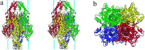
(Quaternary structure of PAL. The four individual monomers are color-coded in red, green, blue, and yellow, displaying the approximate 222 symmetry of the PAL tetramer. (a) Stereoview of PAL from a side perspective. The bracketed areas represent the residu) |
(→Licensing) |
||
| Line 3: | Line 3: | ||
== Licensing == | == Licensing == | ||
{{subst:Non-commercial from license selector}} | {{subst:Non-commercial from license selector}} | ||
| + | |||
| + | Calabrese, Joseph C. et al.<ref name=crystal>http://pubs.acs.org.prox.lib.ncsu.edu/doi/pdfplus/10.1021/bi049053%2B</ref> | ||
Current revision
Summary
Quaternary structure of PAL. The four individual monomers are color-coded in red, green, blue, and yellow, displaying the approximate 222 symmetry of the PAL tetramer. (a) Stereoview of PAL from a side perspective. The bracketed areas represent the residues, arranged in a fan, that are present in PAL and absent in HAL. (b) Top perspective of the PAL tetramer (90° offset from that of panel a). This view looks down into the active sites of two of the subunits; the other two active sites would be seen from a bottom view.
Licensing
{{subst:Non-commercial from license selector}}
Calabrese, Joseph C. et al.[1]
File history
Click on a date/time to view the file as it appeared at that time.
| Date/Time | User | Dimensions | File size | Comment | |
|---|---|---|---|---|---|
| (current) | 13:09, 6 December 2013 | Bryan Toton (Talk | contribs) | 500×173 | 49 KB | Quaternary structure of PAL. The four individual monomers are color-coded in red, green, blue, and yellow, displaying the approximate 222 symmetry of the PAL tetramer. (a) Stereoview of PAL from a side perspective. The bracketed areas represent the residu |
- Edit this file using an external application
See the setup instructions for more information.
Links
The following pages link to this file:
